– Michelle Arnold, DVM (Ruminant Extension Veterinarian, UKVDL)
Johne’s (pronounced Yo-knees) Disease is a chronic disease of severe, watery diarrhea and weight loss in adult cattle caused by the bacterium Mycobacterium avium subsp. paratuberculosis, commonly referred to as “MAP”. These bacteria are very hardy due to a protective cell wall that can withstand harsh conditions and allows survival for long periods in the environment. Once MAP gains entry into an animal, the organism lives permanently within the cells of the large intestine where it multiplies and is then “shed” in the feces in large numbers. Johne’s is a slow, progressive disease that calves pick up around the time they are born but the clinical signs of weight loss and diarrhea do not show up until much later, generally at 2-5 years of age or even older.
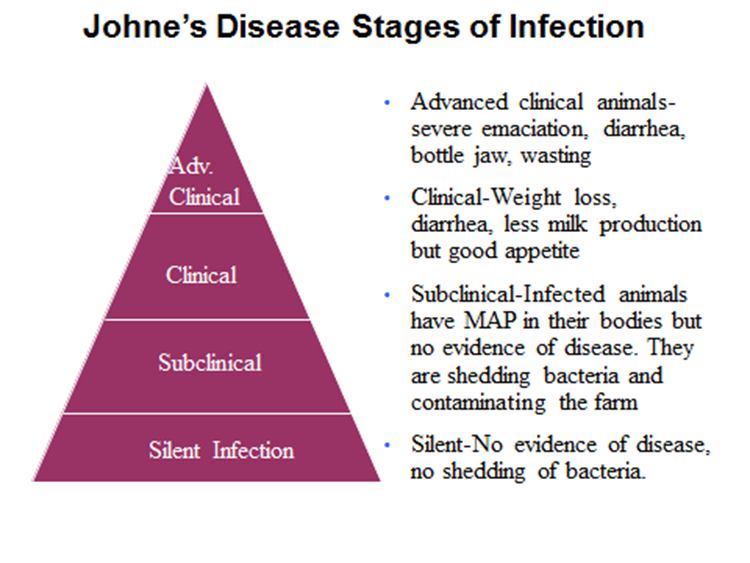
As cow/calf producers, it is easy to buy and sell breeding age animals, especially bulls, with no obvious problems even though they are already infected with the disease. The problem is difficult to detect early in subclinical cattle (subclinical = before symptoms of diarrhea and weight loss develop) but these infected animals often shed high numbers of the MAP organism on the farm. In ideal conditions with moisture and limited sunlight, bacteria can live 8 months in dry feces, 9-12 months in a manure pit/lagoon, 18 months in a water trough, 9-12 months in freezing temperatures and 1 or more years on pasture. This is important because the major route of transmission to newborn calves is nursing teats covered in Johne’s-infected manure. A small number of calves may get the disease while still in the uterus of an infected cow or may ingest the organism from infected colostrum or milk. Once infected, there is a long incubation period (2-7 years) then the disease begins its progression from a silent stage to an advanced disease stage. No effective treatment is available.
In a typical herd, for every animal in the advanced or clinical stage of disease, there are often many other cattle in earlier stages of the disease. Control measures center upon preventing exposure of susceptible animals to the infectious agent, identifying and eliminating infected animals from the herd, and preventing entry of infected animals into the herd. With early diagnosis and prevention of spread, Johne’s Disease will not develop into a significant herd problem five to ten years in the future. Buyers of breeding livestock should make every effort to purchase animals that are not MAP infected. Similarly, seedstock producers should anticipate this request and establish a routine of testing and culling any cattle that test positive for the organism. Seedstock herd owners are commonly reluctant to test for Johne’s Disease for fear that a positive diagnosis will ruin their reputation. However, a herd’s reputation may be damaged much more severely by selling a MAP-infected animal to a customer and introducing this contagious, incurable disease into the buyer’s herd. The US Voluntary Bovine Johne’s Disease Control Program specifies the testing requirements to officially classify a herd from Test Negative Level 1 (lowest) up to Level 6 (best). The more years of testing following this consistent regimen will yield greater confidence and knowledge of the true Johne’s status of the herd.

Picture from W.C. Davis, S.A. Madsen-Bouterse, Veterinary Immunology and Immunopathology 145 (2012) 1– 6
So how do you begin? A screening test of all animals at least 2 years of age, such as the Johne’s ELISA test for antibodies in serum (blood), is rapid and low cost but not 100% accurate. Any positive animals on ELISA should be confirmed by detection of the MAP organism in the feces by polymerase chain reaction (PCR). Both of these tests are available at the UK Veterinary Diagnostic Laboratory (for more information, visit our website: vdl.uky.edu). Animals found positive should be removed from the herd promptly. Testing and culling over multiple years along with good herd management will lead to zero or low MAP test prevalence in your herd. Contact your local veterinarian to find out more about the control and prevention of Johne’s Disease.
Picture E is an endoscopic view of normal cow intestine. Picture F is an endoscopic view of a Johne’s infected cow, showing the characteristic swelling and corrugation of the lining of the intestine that occurs at the clinical stage of infection. Thickening of the intestinal wall results in poor absorption of nutrients and diarrhea in affected animals.