Explaining Science – vermiform mites

You have heard of mites – minute arachnids that have four pairs of legs when adult, are related to the ticks and live in the soil, though some are parasitic on plants or animals. But what are vermiform mites? Maybe you have heard of vermi-compost, a composting technique that uses worms (like your earthworm in the garden) to decompose organic matter. So vermiform mites are mites with a body shape like a worm:

worm-shaped nematalycid Osperalycus

Why are they shaped like a worm, you may ask – To find out more I interviewed Samuel Bolton, former PhD student in the acarology collection at our museum, now Curator of Mites at the Florida State Collection of Arthropods. Sam’s main research interest is in mites that live on plants and in the soil, especially Endeostigmata, a very ancient group of mites that dates back around 400 million years, before there were any trees or forests. Sam’s PhD research with Dr. Hans Klompen here at OSU, was focused on a small family (only five described species) of worm-like mites, called Nematalycidae.

side note: You may have heard of Sam’s research in 2014 when he discovered a new species of mite, not in a far-away country, but across the road from his work place in the museum.

When Sam started his research it was not clear where these worm-like mites in the family Nematalycidae belong in the tree of life. To find out Sam studied several morphological characters of Nematalycidae and other mites. He focused in particular on the mouth-parts of this group. As he learned more about the mouth-parts of this family, he found evidence that they are closely related to another lineage of worm-like mites, the gall mites (Eriophyoidea). Eriophyoidea have a sheath that wraps up a large bundle of stylets. They use these stylets to pierce plant cells, inject saliva into them and suck cell sap.
Although Nematalycidae don’t have stylets, one genus has a very rudimentary type of sheath that extends around part of the pincer-like structures that have been modified into stylets in Eriophyoidea.

So what did Sam and his co-authors discover?

“.. Not only are gall mites the closest related group to Nematalycidae, but the results of our phylogenetic analysis places them within Nematalycidae. This suggests that gall mites are an unusual group of nematalycids that have adapted to feeding and living on plants. Gall mites use their worm-like body in a completely different way from Nematalycidae, which live in deep soil. But both lineages appear to use their worm-like bodies to move around in confined spaces: gall mites can live in the confined spaces in galls, under the epidermis (skin), and in between densely packed trichomes on the surface of leaves;  Nematalycidae live in the tight spaces between the densely packed mineral particles deep in the soil.”

This research potentially increases the size of Sam’s family of expertise, Nematalycidae, from 5 species to 5,000 species. We have yet to confirm this discovery, but it is highly likely that gall mites are closely related to Nematalycidae, even if they are not descended from Nematalycidae. This is interesting because it shows that the worm-like body form evolved less frequently than we thought. This discovery also provides an interesting clue about how gall mites may have originated to become parasites. They may have started out in deep soil as highly elongated mites. When they began feeding on plants, they may have used their worm-shaped bodies to live underneath the epidermis of plants. As they diversified, many of them became shorter and more compact in body shape.

I wish I could tell you now to go out and look for these oddly shaped mites yourself, but you really need a microscope. Eriophyoid mites are minute, averaging 100 to 500 μm in length. For your reference, an average human hair has a diameter of 100 microns.

eriophyoid Aceria anthocoptes

Reference:

Bolton, S. J., Chetverikov, P. E., & Klompen, H. (2017). Morphological support for a clade comprising two vermiform mite lineages: Eriophyoidea (Acariformes) and Nematalycidae (Acariformes). Systematic and Applied Acarology, 22(8), 1096-1131.


About the Authors: Angelika Nelson, curator of the Borror Laboratory of Bioacoustics, interviewed Samuel Bolton, former PhD graduate student in the OSU Acarology lab, now Curator of Mites at the Florida State Collection of Arthropods, in the Florida Department of Agriculture and Consumer Services’ Division of Plant Industry.


Dynamics of Neo-Tropical Arachnids

Today’s post is a guest post by Andrew Mularo,  an undergraduate student in the Department of Evolution, Ecology and Organismal Biology. He is currently doing his Tropical Behavior Evolution and Ecology research project under Dr. Rachelle M. M. Adams and Dr. Jonathan Shik.

You may love them or you may fear them, but no one can deny the incredible ecological importance of spiders and scorpions. As an aspiring biologist, I have chosen to study the interactions between arachnids and their environment in the tropical rainforests of Panama for the 2017 Tropical Behavioral Evolution and Ecology course. The tropics are a biodiversity hotspot for the majority of the world’s organisms, so there are plenty of creatures to look at. From the smallest spiderling to the largest tarantula, I am curious to see how these cryptic and intriguing animals interact with their ecosystem.

For my project, I am doing an observational study where I am assessing the relationship between leaf litter and arachnid diversity and abundance. I am accomplishing this by creating several 50 meter transects in the Panamanian rainforest, sampling leaf litter with 1 square meter quadrants along each transect. For each quadrant, I take a measurement of leaf litter depth, and sift through the leaves to extract any organisms out of the area. Back at the lab, I sort through the organisms, first finding any arachnids in the sample, and then any other insect or invertebrate, such as ants, beetles, millipedes, snails, mites and many others. With these data, I hope to make a correlation between leaf litter abundance and arachnid diversity and abundance, as well as a correlation between the diversity of potential prey items and arachnid predators.

Naturally, the majority of the organisms that I have been assessing have been very small, from the size of a thumbnail to not even being visible to the human eye. However, there

Wandering Spider (Photo by A. Mularo)

are several occasions where I have observed some extremely imposing arachnids in the tropical forest. One of these includes the huntsman spider, an extremely large nocturnal species that does not rely on a web to capture its prey. This family of spiders is very poorly researched, and is largely unknown how dangerous the venom is for the majority of species. However, they are quite shy, and often scurry away at the sight or sound of a human.

Another fascinating group of organisms I see occasionally are scorpions. The two pictured below are from the genus Tityus, whose venom is very potent. I found the two in the picture below, which we believe to be different species, huddled in close quarters in the water well of a bromeliad. While potentially dangerous, these are a relatively uncommon sight in the rainforest. Nevertheless, it is always good to be careful where you step.

Tityus scorpions (photo by A. Mularo)

While many of them are feared, arachnids are some of the most fascinating organisms on the planet. They come in all shapes and sizes, and have a wide array of interesting characteristics that are of great interest to scientists. Being interested in biology since I was a child, I have always dreamed of coming to the tropics so I could study the vast diversity of organisms, and I could not have picked a better group of organisms to focus on!

Our big day is tomorrow

Tomorrow, Saturday April 22, from 10 AM – 4 PM we will open our doors and welcome all of you to visit our hidden treasures in the natural history collections of The Ohio State University. Stop by and talk to the curators who meticulously keep these specimens and make them available to students and researchers for study throughout the year. This is your chance each year to see what we do and to support our efforts.

The event is FREE and so is parking. We will have many activities for children including face painting, the very popular bugs-in-goo, a live arthropod zoo … and this year new, for anyone over 15 years, guided sessions on scientific illustration, drawing natural history specimens.

Enjoy some photos from last year events

This slideshow requires JavaScript.

The set-up for tomorrow is in full swing, here is what I have seen so far

This slideshow requires JavaScript.


About the Author: Angelika Nelson is curator of the Borror Laboratory of Bioacoustics and coordinates social media and outreach at the museum.

*** We hope to see you tomorrow ***

Ticks in pictures

Some more about ticks.  No, not The Tick comic or the movie Ticks … both may be entertaining, but they feature completely inaccurate depictions of ticks.

Let’s talk about real ticks:  Ticks are rather large mites. To demonstrate this, here is a family portrait:

family portrait of Ixodes pacificus, California Dept. of Public Health

Family portrait of Ixodes pacificus, California Dept. of Public Health [public domain]

From left to right, larva (6 legs), nymph (8 legs), male and female of Ixodes pacificus, the Western black-legged tick, from the west coast (you can see them with the naked eye, therefore they are big).

All members of the family feed on host blood using highly modified mouthparts, but only larvae, nymphs, and females engorge (feed to the point where their body truly swells up).

close-up of mouth parts of Amblyomma extraoculatum, U.S. National Tick Collection (USNMENT00956315)

Close-up of mouth parts of Amblyomma extraoculatum, U.S. National Tick Collection (USNMENT00956315)

Here are some nice examples of engorged females.  Keep in mind that while engorged ticks are easy to find, they are often difficult to identify.

Most of the ticks we encounter in Ohio have females that feed only once.  They engorge, convert all that host blood into a single mass of hundreds to thousands of eggs, and die.

tick with eggs (c) Univ. Nebraska, Dept. Entomology

Tick with eggs, Univ. Nebraska, Dept. Entomology

Ticks in general get really bad press.  Kind of sad, because ticks are very good at quite a few things, like surviving (some can survive hours under water or years without food), or manipulating your immune system (using a dizzying array of chemicals often found only in ticks). On second thought, that may not strike most people as positive, so let me end with a few pictures of beautiful creatures. I already introduced Amblyomma americanum, which occurs in Ohio, the others are African, A. chabaudi on tortoises in Madagascar, A. variegatum usually on cattle. Amblyomma variegatum is the main vector of heartwater, a disease making cattle herding impossible in parts of Africa, but still, very pretty.

This slideshow requires JavaScript.

See some more of these specimens close-up, but at a safe distance through microscopes at our Annual Open House, April 22, 2017.


Dr. Hans Klompen, Professor EEOBiology at OSUAbout the Author: Dr. Hans Klompen is professor in the department of Evolution, Ecology and Organismal Biology and director of the Ohio State University Acarology Collection.

*** Which of these ticks is your “favorite”? Let us know on Facebook ***


Know your ticks: Ohio

Daffodils are in bloom, students walk around in shorts and T-shirts, so it must be the beginning of tick season.  And indeed, the first ticks are out and questing (= searching for a host). This might be a good time to talk about ticks in Ohio.  Ohio is not a major center for tick diversity, but it has some diversity.  Most people only know the three main people biters, Dermacentor variabilis (American dog tick), Amblyomma americanum (lone star tick), and Ixodes scapularis (deer tick), so let’s start with these:

Dermacentor variabilis is perhaps the most widespread and common tick in Ohio.  Immatures feed on rodents and other small animals, but adults feed on medium (opossums, raccoons, dogs) to large (humans) mammals.  Of the “big three” this species is the most tolerant of drying out, and the most likely to be encountered in open areas.  The main activity period for adults is mid-April – mid-July.  D. variabilis is the vector of, among others, Rocky Mountain Spotted Fever (RMSF) and tularemia.  Columbus used to be a focal area for RMSF, but the disease is less common now.  D. variabilis may also cause tick paralysis, although less frequently than the related D. andersoni from the Rocky Mountains region.

American dog tick

Dermacentor variabilis American dog tick

Amblyomma americanum used to be uncommon in southern Ohio, but has increased in numbers and range over the last decades.  This is part of a general trend.  In the eastern U.S., this species is rapidly expanding its range northwards.  All instars, larva, nymph, and adult feed on mid-size to large animals, incl. humans.  Like D. variabilis, females can deposit very large clutches of eggs, but in this case the resulting larvae often stay together.  If you are unlucky and step close to a mass of these “seed ticks”, you may be attacked by hundreds of ticks simultaneously.  These ticks are active in all warm months of the year.  Unlike D. variabilis, “Lone stars” are not common in open areas, preferring more shady and humid sites.  For a long time A. americanum was listed as vectoring few human diseases, but it has now been identified as vector of human monocytic ehrlichiosis and STARI, and possibly tularemia and Q-fever.

lone star tick

Amblyoma americanum lone star tick

Ixodes scapularis appears to be an even more recent resident.  This species was rare or absent in Ohio before 2010, but has now been found in a majority of Ohio counties.  The reason for this sudden expansion is unclear.  This is a relatively small species.  Larvae can be found in summer, nymphs late summer, and adults in fall and early spring.  Immatures tend to feed on smaller sized hosts, e.g. rodents, small birds, while adults prefer larger hosts, such as deer.  However, all instars may attach to humans.  Nymphs are considered the most problematic: they are small (thus often undetected), and can be infected with e.g. Lyme disease (unlike the even smaller larvae).  Like A. americanum, this species prefers shady, humid environments.  New subdivisions build in forests, resulting in large amounts of forest edges with lots of deer, have been a very good habitat for this tick in New England.  Ixodes scapularis has become famous as the vector for, among others, Lyme disease, human granulocytic anaplasmosis, and babesiosis.  Co-infection is common in New England and appears to result in increased pathology.

deer tick

Ixodes scapularis deer tick

So much for the common people biters.  It is important to note that most species of tick rarely if ever bite people.  They prefer different, usually smaller, hosts.  For example, Rhipicephalus sanguineus, the brown dog tick prefers feeding on dogs.  It is one of the few species that may occur indoors in dog kennels etc.  Haemaphylis leporispalustris appears to be specialized on hares and rabbits.  Several Ixodes species, I. cookei, I. dentatus, I. kingi, I. marxi, can be found on small to medium sized mammals, often associated with nests or burrows.  Finally, the so-called soft ticks, family Argasidae, are represented by only a single species in Ohio, Carios kelleyi, primarily found in bat colonies.

Find out more about the ticks’ life cycles and their diseases.

Dr. Hans Klompen, Professor EEOBiology at OSUAbout the Author: Dr. Hans Klompen is professor in the department of Evolution, Ecology and Organismal Biology and director of the Ohio State University Acarology Collection.


*** Have you found a tick yet this spring? send us a photo of your specimen on Facebook! ***


Everyday Life at the Museum-part 1

Visitors to our Annual Open House – by the way, the next Open House is coming up soon on Saturday April 22, 2017 – often wonder what everyday life in our museum collections looks like. During the Open House we showcase specimens in fabulous displays but how do we accession and maintain specimens throughout the year? To find out I took a walk through our building on a weekday morning and stopped by each collection to get a snapshot of our students’ and staff’ workdays. Watch the short videos below to get some behind the scenes insights and see how important the help of OSU undergraduate students is for our collections.

In the Borror Laboratory of Bioacoustics Evolution and Ecology major Morgan VanDeCarr digitizes recordings of House Finches Haemorhous mexicanus. The song of these birds was recorded onto a reel-to-reel tape by researcher Erik Bitterbaum at Occidental College in 1976.

In the Triplehorn insect collection Art major Katherine Beigel takes images of tiny insect specimens under a microscope and stacks them into one composite image using special software. Here she shows us a Coleoptera, beetle specimen.

In the Acarology collection Dr. Hans Klompen, Professor in the OSU department of Evolution, Ecology & Organismal Biology, talks to a student. The tick and mite specimens are neatly shelved and ready for the next Acarology Summer Program (June 19 – July 7).

In the Tetrapods collection Evolution and Ecology major Chelsea Hothem updates location information in the computer database. To get accurate data she often goes back to the specimens and reads information on the tags.

We will look behind the scenes in the herbarium, the mollusc and the fish collection on Friday!


About the Author: Angelika Nelson is curator of the Borror Laboratory of Bioacoustics and the museum’s social media and outreach manager.


*** Do you have any questions? We would be happy to answer them ***


More about the biology of a parasitoid mite

Following Monday’s blog post, we continue to explore the life of  Macrodinychus mites that parasitize an invasive ant species in Mexico, the Longhorn crazy ant. Today Dr. Hans Klompen shares some of the details of the mite’s life cycle that he has discovered with us.

Here is an image of two Macrodinychus larvae that were found attached to an ant pupa. We had to magnify the ant 400 times to make the mites visible. The larvae are tiny, even in mite-standards, while the adults are large, 1 mm or more in length.

What do the larvae and nymphs look like?


Tell us a little bit more about the biology of these mites, e.g. how does the female give birth to young?


How do the mites disperse to new hosts?


Why is this research important?


What do you think?  Can/should these mites be used to control invasive ant species?
We would like to hear from you – please leave a comment.

*******************************************************

Reference:

Image of mite larvae on appendages and gravid female from
Krantz, G. W., Gómez, L. A., González, V. E., & Morales-Malacara, J. B. (2007). Parasitism in the Uropodina: a case history from Colombia. In Acarology XI: Proceedings of the International Congress (pp. 29-38).

other images from
Lachaud, J. P., Klompen, H., & Pérez-Lachaud, G. (2016). Macrodinychus mites as parasitoids of invasive ants: an overlooked parasitic association. Scientific Reports, 6.
Dr. Hans Klompen, Professor EEOBiology at OSUAbout the Author: Dr. Hans Klompen is professor in the department of Evolution, Ecology and Organismal Biology and director of the Ohio State University Acarology Collection.

Mites as parasitoids of invasive ants

Another post in our series Explaining Science – bringing scientific discoveries focused around biodiversity to your living room.


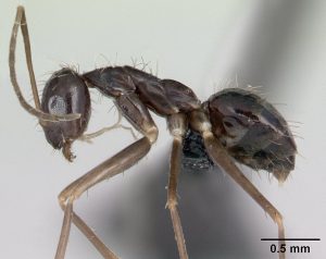
Ants are fascinating creatures, often living in large colonies. Some of you may be familiar with this behavior as a  nuisance in your home, e.g. with carpenter ants or fire ants. Ant biology and their way of social living fascinates researchers, and some of the ants’ behavior may be quite similar to what we see in our societies. Just recently a study reported how Gene-Modified Ants Shed Light on How Societies Are Organized. But did you know that these small ants can themselves become hosts for even smaller animals? Mites, in particular species in the genus Macrodinychus, have evolved to parasitize ants. They feast on the content of ant pupae, the larval stages of ants, to nourish their own development. “Vampire mites” is what Dr. Hans Klompen, acarologist and Professor in the department of Evolution, Ecology and Organismal Biology, calls them. By the way, the ant species these mites parasitize is called Longhorn crazy ant, an invasive ant species with a cool name.

Listen to an interview with Dr. Klompen about his recent publication in Scientific Reports “Macrodinychus mites as parasitoids of invasive ants: an overlooked parasitic association” and learn about “a bizarre little group of mites”” that he studies.

How does one find out about mites, often microscopical creatures, living on ants, in particular when you are a researcher based in Ohio while the ants live mainly in the tropics?

One needs good collaborators at El Colegio de la Frontera Sur (ECOSUR), Gabriela Perez-Lachaud and Jean-Paul Lachaud, who study ants and noticed mites parasitizing their study subjects.

How did the project of describing a new mite species evolve into more?


Macrodinychus multispinosus Sellnick larva

Macrodinychus multispinosus Sellnick larva

How often do these mites attack ants and which species of ants?

Longhorn crazy ant

Longhorn crazy ant, the host
(c) The photographer and www.antweb.org, CC BY-SA 3.0


Do the mites attack all different colonies of ants?


So what do we know about the life history of this mite whose developmental stages, its nymphs, feast on ant pupae? Find out more results from Dr. Klompen’s research on these mites in Friday’s post!

***************************************************************

Terms you may want to familiarize yourself with:

Mites are small arthropods, closely related spiders and scorpions, with two body regions, no antennae, and four pairs of legs as adults.

The life cycle of these mites is  composed of five active stages: egg, larva, protonymph, deutonymph, and adult

ventral – the underside of an animal, the belly

dorsal – the upper side of an animal, the back


Reference:

Lachaud, J. P., Klompen, H., & Pérez-Lachaud, G. (2016). Macrodinychus mites as parasitoids of invasive ants: an overlooked parasitic association. Scientific Reports, 6.
Dr. Hans Klompen, Professor EEOBiology at OSUAbout the Author: Dr. Hans Klompen is professor in the department of Evolution, Ecology and Organismal Biology and director of the Ohio State University Acarology Collection.
 *** We would like to hear from you – please leave a comment ***

Itchy noses – a perfect ecosystem for mites & ticks?

Mites on occasion have become extreme specialists in selecting the places where they live. Take the noses of vertebrates. It may not seem much, but a wide variety of mites call it home. Mites can do different things while in the nose. Rhinoseius and some Proctolaelaps species use hummingbird noses to move from flower to flower.

hummingbird sticking bill into red flower

Hummingbird sticking its “nose” into a flower (did you know that the nostrils of birds are located at the base of the bill?)

The mites race up or down the bill when the bird is feeding to get in or out of the nose as they move between flowers. Nice and fast transportation but it can be tricky. If a male ends up in a flower already occupied by males of a different species they may get attacked and killed. As always make sure you get off at the right bus stop.

Dispersal is also the goal for some Halarachnidae living in seals. They live most of their lives in the lungs, but larvae will crawl up into the nose and get dispersed by sneezing. It is not sure whether they irritate the nose and make the seals sneeze or whether they just take advantage of seal sneezing.  This form of dispersal is of course a bit random.  For example, a paper from 1985 described a case where an ophthalmologist recovered a halarachnid mite from the eye of a patient with severe eye discomfort.  The man had been watching the walrus exhibit at Sea World.  Moral of the story: be aware of flying debris when visiting the seal exhibit.

Most nose-inhabiting mites are true parasites. Some chiggers (Trombiculidae) are found only in noses. So do most species of Gastronyssidae, although I have collected some skating around on the eyeball of fruitbats, and 1-2 others appear exclusive to the stomach of such bats. In birds we sometimes see a split in microhabitat: Rhinonyssidae live in the slimy parts of the nostrils, Ereynetidae skate on top of the slime, and Turbinoptidae live in the dryer section further down.

rhinonyssid mite from nose of pigeon

Rhinonyssid mite from nose of pigeon

Noses are true ecosystems.


About the Author: Dr. Hans Klompen is professor in the department of Evolution, Ecology and Organismal Biology and director of the Ohio State University Acarology Collection.


Reference:

Webb, J.P., Jr., Furman, D.P. & Wang, S. (1985) A unique case of human ophthalmic acariasis caused by Orthohalarachne attenuata (Banks, 1910) (Acari: Halarachnidae). Journal of Parasitology, 71 (3), 388-389.

Mite art

The Berlese Alphabet

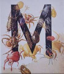
These letters are from the four-volume “Acaroteca” of Antonio Berlese, (1863-1927) in which he maintained records of his named specimens. Berlese illustrated this catalogue with a large letter at the beginning of each section. The mites are examples of those whose genus name begins with that letter. Not all letters were completed before his death, and several pages have been heavily stained.

Norton, 2008

The Berlese Alphabet

Following up on unusual ways to treat (or image) mites, here is an example that is both beautiful and very traditional, following the medieval tradition of illuminated letters. This image is of a poster assembled by Roy Norton featuring most of the letters of the Berlese alphabet. In case you have never heard of Berlese, Antonio Berlese was an Italian entomologist (1863 – 1927) who studied agricultural pest insects. He put together a catalogue of his collection of mites, referred to as the Catalogue of the Berlese Acaroteca. He included about 1600 species, the entries are arranged alphabetically by species according to Berlese’s final specific and generic concept. Each section begins with a large letter featuring a mite whose genus name begins with that letter.

letter M with images of mites from the Berlese alphabet

Letter M from the Berlese Alphabet, © Cal Welbourn

The individual drawings are simply gorgeous, and most specimens can easily be identified to genus. Of course many generic names have changed since early 20th century, so I would not recommend this as a work of taxonomy, but the Berlese alphabet remains a great work of “mite art”.


About the Author: Dr. Hans Klompen is professor in the department of Evolution, Ecology and Organismal Biology and director of the Ohio State University Acarology Collection.