Strange things to do with ticks

When most of us think of natural history collections we see well-labeled, nicely arranged rows of jars, sets of herbarium sheets, or pinned insects, and this is certainly a curator’s ideal. But this does not acknowledge the occasional outburst of mis-applied creativity leading to a novel approach to preserving specimens. Every collection probably has a few examples, and I thought I should share some from the acarology collection. The collection includes some ticks processed in ways that are simultaneously novel, creative, and useless.

The standard way to preserve ticks is in fluid, mostly 70-95% ethanol. Ideally in good vials with complete labels, and a barcode linked to a properly functioning database.

vial of ticks (Ixodes lemuris) with proper labeling

vial of ticks (Ixodes lemuris) with proper labeling

One less than great alternative is diluted formalin, at least for a brief period of time. This mixture does not evaporate as fast as ethanol (good in warm regions and in the absence of good containers) but it diminishes the value of the specimens because it destroys DNA. Still, this is not very odd. “Weird” would be one word for the option of pinning ticks.

pinned and slightly shriveled ticks

pinned and slightly shriveled ticks


We have a few of these in the OSU Acarology Collection. These specimens are essentially useless. Insects, with their hard cuticles, do quite well on pins, but generally soft-bodied organisms like ticks just shrink and shrivel, in the process destroying many valuable characters.


While tick adults and (usually) nymphs are fluid preserved, tick larvae are small enough that they can also be put on slides. A well-prepared slide of an unengorged tick larva can be a thing of beauty.

slide of larval Haemaphysalis lemuris

slide of larval Haemaphysalis lemuris

Under a microscope you can observe very fine detail of the cuticle structure, leg hairs, mouthparts, etc. Of course not everybody is as narrow-minded as the above lines suggest. For example, one specimen of an engorged adult dog tick (Dermacentor variabilis) in our collection shows a reckless disregard of the rule that slide mounting is only for larvae. The specimen is encased in a 2mm high wooden “box” placed on a slide, filled with mounting medium and topped with a glass cover slip.

side view of slide with female Dermacentor variabilis

side view of slide with female Dermacentor variabilis

The overall resulting structure is far too thick and too opaque to be usable, but the craftsmanship exhibited in making this “box” can only be described as exquisite.


I would like to close with a salute to those among us that are not bound by conventions and that bravely go where nobody has gone before. Just don’t do it again.


About the Author: Dr. Hans Klompen is professor in the department of Evolution, Ecology and Organismal Biology and director of the Ohio State University Acarology Collection.

Mites and moths

Following some earlier blogs about recently acquired collections I present to you here the Treat collection. This collection was assembled by Asher E. Treat a researcher at City University of New York and the American Museum of Natural History, also New York. This collection is one of the best in the world for mites associated with Lepidoptera (butterflies and moths). Mites have been found associated with most terrestrial and many aquatic organisms, but when it comes to insect hosts, mites on Coleoptera (beetles) and Hymenoptera (bees and wasps) are clearly the most numerous, diverse, and well-known. Still, Lepidoptera have a variety of associated mites.

The Acarology Collection acquired this collection 4 years ago, some years after Treat’s death. The collection consisted of about 37 slide boxes of exceptionally well labelled microscope slides and half a dozen insect drawers of pinned moths (all labelled as hosts of specific mite specimens). The Lepidoptera are being processed at the Triplehorn Insect collection, while we, the Acarology collection, have been working on processing (mostly databasing) the slides. This is proving to be a major job.


Image of a female of Dicrocheles phalaenodectes, the moth ear mite

Image of a female of Dicrocheles phalaenodectes, the moth ear mite

Treat got interested in mites associated with moths after finding mites in the ears of noctuid moths. In the process, he figured out the quite amazing life histories of some mites associated with these moths. The most famous is Dicrocheles phalaenodectes, the moth ear mite (family Laelapidae).

These mites break through the tympanic membrane of the ear of the moth and form small colonies inside the ear. By itself not too surprising, but the interesting part Treat discovered was that these mites are always found in one ear only, rarely if ever in both ears. In a way this makes sense. By breaking the tympanic membrane the mites make the moth deaf in that ear. Moths need their hearing to avoid predators (for example bats) so a deaf moth would be easy prey. However, a moth with one functional ear is still able to avoid bats, perhaps not as well as if it had two functional ears, but close enough. Which leaves the question: how do the mites manage to limit infestations to one ear?

Treat did many careful observations and follow-up experiments on this aspect and found that the mites have a very specific set of behaviors ensuring only one ear will be parasitized. The first female to get on a moth (nearly always a fertilized female, the immatures and males do not colonize) crawls to the dorsal part of the thorax, explores a little, after which she proceeds to one ear. Any future colonizers will first go to that same dorsal part of the thorax of the moth and follow the initial female to the same ear. It appears the mites lay a pheromone trail that guides newcomers to the already infested ear, and away from the uninfested one.

Drawing of relative positions of mites in a moth ear

Drawing of relative positions of mites in a moth ear

To complete the cycle, young females leaving the ear initially wander around the hosts body (mostly the thorax), congregating around the head at night. They leave the moth by running down its proboscis when it is feeding on flowers. On the flowers, the mites wait for their next host.
Another mite family that is specialized on Lepidoptera, the Otopheidomenidae, is also parasitic, and they will also show up near the ears, but they do not pierce the tympanic membrane, so they do not cause deafness. Unfortunately, we know much less about them, Treat was never able to study their behavior. A range of other mite families have representatives that are regularly found on Lepidoptera, but they are not specialists at the family level: Ameroseiidae, Melicharidae, Erythraeidae, Iolinidae, Cheyletidae, Acaridae, Carpoglyphidae, and Histiostomatidae. That list excludes the occasional “vagrants” that can be found on moths, but that are unlikely to be living on them for extended periods of time. All in all, quite a diverse community.
For those interested in knowing more, Treat wrote a book “Mites of Moths and Butterflies” (1975, Cornell University Press) that is a rare combination of good scholarship (especially natural history) and readability.

Title page of Treat's book on moth mites

Title page of Treat’s book on moth mites

Treat was very careful and noted things like host specimen numbers (if available), which allows current researchers to track down the exact moth from which a given mite came.

This is currently a common approach, but Treat started this in the 1950-ies. And there is more. Based on Treat’s label data we know not only the name of the hosts and the specific locality, but also gender of the host, whether the left or right ear was infested, and the exact part of the body the mites were found on. So we have excellent information, directly from the slides, showing that Proctolaelaps species (family Melicharidae) are nearly always found near the base of the palps [as an aside, Proctolaelaps is a bit of an unfortunate generic name, combining “procto-” = anus and “laelaps” = hurricane; presumably the name

Microscope slide from the Treat collection

Slide from the Treat collection

refers to a relatively large anal shield]. Such complete data are fantastic for future research, but they also mean a lot more work processing these slides, as every slide has lots of unique data. I want to thank George Keeney, part-time curator of the acarology collection and a series of volunteers, Ben Carey, Rachel Hitt, Mitchell Maynard, Ben Mooney, Jake Waltermyer, and Elijah Williams, for their hard work in accessioning this material.


About the Author: Dr. Hans Klompen is professor in the department of Evolution, Ecology and Organismal Biology and director of the Ohio State University Acarology Collection.

Smile!! Imaging in the Acarology collection


Velvet mite, <i>Leptus</i> sp. (photo Rich Bradley)

Velvet mite, Leptus sp. (photo Rich Bradley)

Working with small organisms such as mites presents some interesting problems.  One of those is getting folks interested in just thinking about something they usually cannot see.  Of course a few mites are big enough to see and critters like ticks, velvet mites and water mites do enjoy some “popularity”.  But for most mites we have only fairly bad photographs of crushed specimens on slides (often good for seeing specific characters, but horrible for outreach).  Without nice images, we can still go for gross (I do admit that I occasionally enjoy going that route), with disgusting images of humans with scabies (Sarcoptes scabiei), or sad looking plants covered in spider mite silk, but it is not the same.  And it really does not do much for improving the image of mites.  Plus, most mites do not do any significant damage.  So how should we present the “real” mites?

Let’s start with the high-tech approach.

Scanning electron microscopy (SEM) was the first good option.  Because of the high depth of field, SEM gives very nice, 3-D images at very high magnification.  It works well for highly sclerotized mites (oribatid adults, uropodids) but it is less successful with small, soft-bodied species because most SEM techniques require critical point drying, which makes those soft bodied mites shrivel into ugly blobs.  Using live mites can alleviate that problem but (a) most SEM operators do not like it (it introduces humidity into the machine and may damage it) and (b) mites are often not cooperative.  As a beginning graduate student I had the option of putting some live sarcoptid mites in an SEM.  The specimens were on sticky tape and everything looked great as the SEM got close to vacuum.  However – there is always a “however” in these stories – just before we could get our first images the last of the great looking mites managed one more spurt of energy, and fell over, exposing nothing but its sticky stuff covered underside.  That took care of my hopes for great pictures.

Leg of <i>Osperalycus tenerphagus</i>, LT-SEM

Leg of Osperalycus tenerphagus, LT-SEM, colorized image

Low Temperature SEM (LT-SEM), uses liquid nitrogen to flash freeze the mites (no critical point drying), allowing views of live mites frozen in time.  This completely bypasses the shriveling problems of standard SEM with stunning results.  The folks at the USDA in Beltsville, MD, have generated some beautiful images of plant feeding mites.  Sam Bolton, graduate student in acarology, worked with them to get the images of his “dragon mite”.  Add some color (based on images of the mite taken before going into the SEM) and we end up with poster-worthy pictures.

Even LT-SEM has limitations, as we can only see surface structures.  We are overcoming that limitation by using confocal laser scanning microscopy.  With this technique you can also see internal structures, and with the right software, you can construct 3-D images that can be sliced any way you wish.  Not quite as detailed as Synchroton X-ray microtomography, but that technique requires a particle accelerator the size of a football field, and at this point only one (in Grenoble, France) seems to be set up to handle images of mites.  Confocal microscopy is great for research, but 3-D printing of the models also makes for excellent teaching tools (or gift store items).

A Confocal 3-D model of <i>Daidalotarsonemus</i> sp.  B 3-D print of the same

A Confocal 3-D model of Daidalotarsonemus sp. B 3-D print of the same

Of course all of those techniques require highly specialized and expensive equipment.  For day-to-day use we use an automated compound microscope and image stacking software to generate images that are both relatively detailed and retain some color.  These tools finally allow generation of well-focused detailed images of very small organisms and structures.  Unfortunately we do lose some of the 3-D effects in the process of image stacking, but on a good day, these images are publication quality.

For more hands-on displays, mites can be viewed on a 40 inch TV-screen using a video camera connected to a compound or dissecting microscope. This is wonderful for teaching (6-12 people can all watch what you are seeing in the microscope), research (sometimes the image on the TV is more detailed than the one you get through the eyepieces of the microscope itself), and of course outreach.  We use that set-up at the annual Museum Open House to show live mites in detail.

Mite on TV as used for teaching

Mite on TV

With enough technology, mites are coming out of the deep and dark, and into the light, where they of course belong.  We are slowly generating more high quality images and making them available through the collection database.  Check it out, the small can be beautiful.

About the Author: Dr. Hans Klompen is professor in the Department of Evolution, Ecology and Organismal Biology and Director of the Ohio State University Acarology Collection.


“(S)he got legs,” a look at mite legs


Mites are notorious for not following the “rules.”  For example, we generally teach that you can separate Insects from Arachnids by the number of legs, 3 pairs in insects, 4 pairs in arachnids. Simple, right?  Mites are arachnids, and most have 4 pairs of legs, but not all


And of course legs can get highly modified for various (known or unknown) purposes


About the Author: Dr. Hans Klompen is Professor in the Department of Evolution, Ecology and Organismal Biology & Director of the Ohio State University Acarology Collection.

The case(s) of the missing collection data


Sherlock Holmes silhouettePrevious blog posts have mentioned the value of data on collection dates, geographic localities, hosts (where relevant) and even things like the weather at the time of collection (collecting before or after a rain storm in the desert gives rather different results). For mites, the group I work on, we often want even more detail. Mites can be both very broad in their requirements and very picky. So information on microhabitat can be crucial. For example, in parasitic mites, we would like things like site on a host. If a mite was found in the quill of the secondary wing feathers of a house sparrow, I can probably give a pretty good guess on what it is. Not certain, there are always surprises, but I can make a good first guess. We know this by experience and by examining records in collections that have that level of precision.

In short, life is good as long as there are data, but what if you end up with specimens with little or no interpretable data? In the Acarology collection specimens are usually on microscope slides or in vials with alcohol.

image of good slides with poor data

Good slides with poor data

Many older slides or vials have labels giving only a partial identification plus a number or code, presumably referring to a notebook, letter, or other non-specified source. In principle fine, but without follow-up those notes get lost, misplaced, etc., and that means trouble. In one unfortunate case, roughly 3,000 chigger slides from South Korea, the data were deliberately omitted from slides and vials. These specimens were collected at the end of the Korean war, and it was probably not a good idea to specifically tell everybody where each military base was located. The chiggers were collected at that time and that place to help manage scrub typhus, a disease transmitted by chiggers. Good idea at that time, but it makes recovering the data at this time, sixty years after the armistice, quite tricky.

chigger photograph

A chigger in all its glory

Anyway, so what do you do when you have a bunch of microscope slides or vials with interesting specimens but no, or very little, data? That is where curation becomes a bit of detective work. There are some options.

Before and after microscope labels

Before and after microscope labels

1) If the specimens have at least a code/number, you often can find the same code of the same code format somewhere else in the collection. This is a brute-force method that requires databasing of large numbers of specimens. Luckily, the vast majority of the OSU Acarology Collection is now databased, with most label data captured. Find matching codes, hope that at least one slide has complete information, and if so, you are set for the other slides in the group. Even if the match is not perfect, a similar style of codes may help narrow down where poorly labeled specimens came from. For example, a certain style of code was used for specimens recovered from bats in Costa Rica. Starting with near complete data on 2-3 labels (out of ~400) and using an on-line mammal collection database (VertNet) we recovered all the relevant collection data and even know where the host specimens are located (Los Angeles County Museum in this case).

page from Lipovsky chigger notebook

Page from Lipovsky chigger notebook

2) Miscellaneous field note books, notes, etc. People have asked me why we keep file cabinets worth of old notebooks, notes, and letters from folks that left long ago. Barring proper log books, this may be where the data we want to recover comes from. One good notebook can solve problems with many specimens. Data for many of the Korean chiggers came from a notebook at the bottom of a drawer of largely useless paperwork. Of course it would be nice to have it all digitized, but we do not have the time, people or resources to do that at this time.

3) Sometimes it just takes some luck. For a long time I was going nowhere with a small set of slides with only identifications, and, on one slide, a name that seemed to refer to a locality in India, and something about a dung beetle. Then one day, out of the blue, I got an e-mail from somebody wondering whether I was interested in the remainder of a collection of mites he made from beetles in India. He had already sent me some specimens years ago (my mystery slides) and now I could add some real data. It does not happen very often, but it counts.

As a final note, some of you might think that the above does include quite a lot of interpretation. How accurate are these interpretations? This is a real problem. We try to be conservative in the “guesses” we make, but may fail on occasion. This is why we copy all original label data verbatim and make that available with each specimen. And we have had a few occasions where folks told us our interpretation was wrong. A big thank you to those folks!


About the Author: Dr. Hans Klompen is Professor in the Department of Evolution, Ecology and Organismal Biology & Director of the Ohio State University Acarology Collection. Silhouette of Sherlock Holmes: The Sherlock Holmes Museum. CC Attribution License. http://www.sherlock-holmes.co.uk. All other images courtesy of the author.

Welcome to the OSU Bio Museum blog


Today I have the pleasure to welcome you to OSU Bio Museum, a blog about biodiversity, research and museum work at the Ohio State Museum of Biological Diversity.  This endeavor is the successor to our newsletter. That effort lived in both the physical and digital worlds, but to keep up with the times and changing needs, the blog is a wholly digital enterprise. The purpose remains the same, though: to share with the community the happenings, news, and successes (and sometimes failures) of the Museum. Our plan is to have weekly postings during the academic semester, with the post authors rotating among the different units in the Museum. We will also feature a Media Gallery every week. My objective in this inaugural post is to briefly describe what those units are and how the Museum is organized and functions.

The Museum, let’s call it the MBD for short, coalesced in its present form in 1992 when the University moved the bulk of the biological collections from the Columbus campus to a newly renovated building on West Campus, at our current address of 1315 Kinnear Road.

Museum of Biological Diversity on 1315 Kinnear Road.

Museum of Biological Diversity on 1315 Kinnear Road

For more than 20 years the MBD has been a bit of a strange beast in that it has been a voluntary association among the collections rather than a real, defined administrative unit. Originally, most of the collections were administered by the Departments of Botany, Zoology, and Entomology. Two or three reorganizations later the primary department is Evolution, Ecology & Organismal Biology (EEOB for short) in the College of Arts & Sciences, and a smaller component associated with the Department of Entomology in the College of Food, Agriculture & Environmental Sciences (CFAES). The Entomology connection is a new one as of September 1, 2015, a reflection of a change in my formal appointment to 75% EEOB and 25% Entomology.

Museum IconThe overall mission of the MBD, just as the University as a whole, is teaching, research, and service. Inside the building we have, of course, the collections themselves, but also office and lab space for faculty, graduate students, postdoctoral researchers, emeriti and undergraduate students. The most glaring absence, though, is space dedicated to public exhibits. We compensate for that in two ways, our annual Museum Open House and guided tours of the facility. The tours are organized on an appointment basis only and have encompassed a wide range of groups, from elementary school kids and scout groups to University President’s Club members.  Anyone interested in scheduling a guided tour of the Museum should contact us, or contact one of the collections directly to make arrangements. It’s my personal aspiration that in the future it may be possible to develop exhibit space for the public in the building, but that’s still just a gleam in my eye!

If you have not done so yet, please visit the Museum website and follow our Facebook page.

So far I’ve referred to the units of the MBD without much explanation. What are they? Well, there are seven main collections: the Triplehorn Insect Collection (which I direct, but for which Dr. Luciana Musetti is the real driving force); the Acarology Laboratory (led by Dr. Hans Klompen), the Borror Laboratory of Bioacoustics (led by Dr. Doug Nelson), the Herbarium (Dr. John Freudenstein), the Division of Molluscs (Dr. Tom Watters), the Division of Tetrapods (Ms. Stephanie Malinich), and the Division of Fishes (Dr. Meg Daly). The naming system, as I write this, must seem very confusing – what’s a collection vs. a division? The names are historical artifacts that, perhaps, made some sense at one time, but now they’re all basically equivalent. As you’ll see in my descriptions below and in future posts, there is a lot of variation among collections in their size, staffing, history and aspirations. So let’s go through the seven units:

Triplehorn collection icon, genus NeomidaCharles A. Triplehorn Insect Collection. The insect collection contains about 4 million prepared specimens, nearly 3,000 primary types, and one of the world’s largest leafhopper collections. The collection formally began in 1934 by Prof. Josef N. Knull, and has strong holdings in beetles (Coleoptera), Hemiptera (true bugs and hoppers), Hymenoptera (ants, bees, wasps), Odonata (dragon- and damselflies) and Orthoptera (grasshoppers and crickets). Originally the specimens largely came from the United States, but we have expanded significantly since then. Recent collecting trips have been made to Brazil, South Africa, Australia, and Malaysia (Sarawak). Ongoing research is focused on the systematics of parasitic wasps and the development of information technologies to share specimen data and images globally.

To know more about the Triplehorn collection, visit the website and follow the collection’s lively social media presence, which include the Pinning Block blog, a Facebook page, a Flickr image site & a Twitter feed.
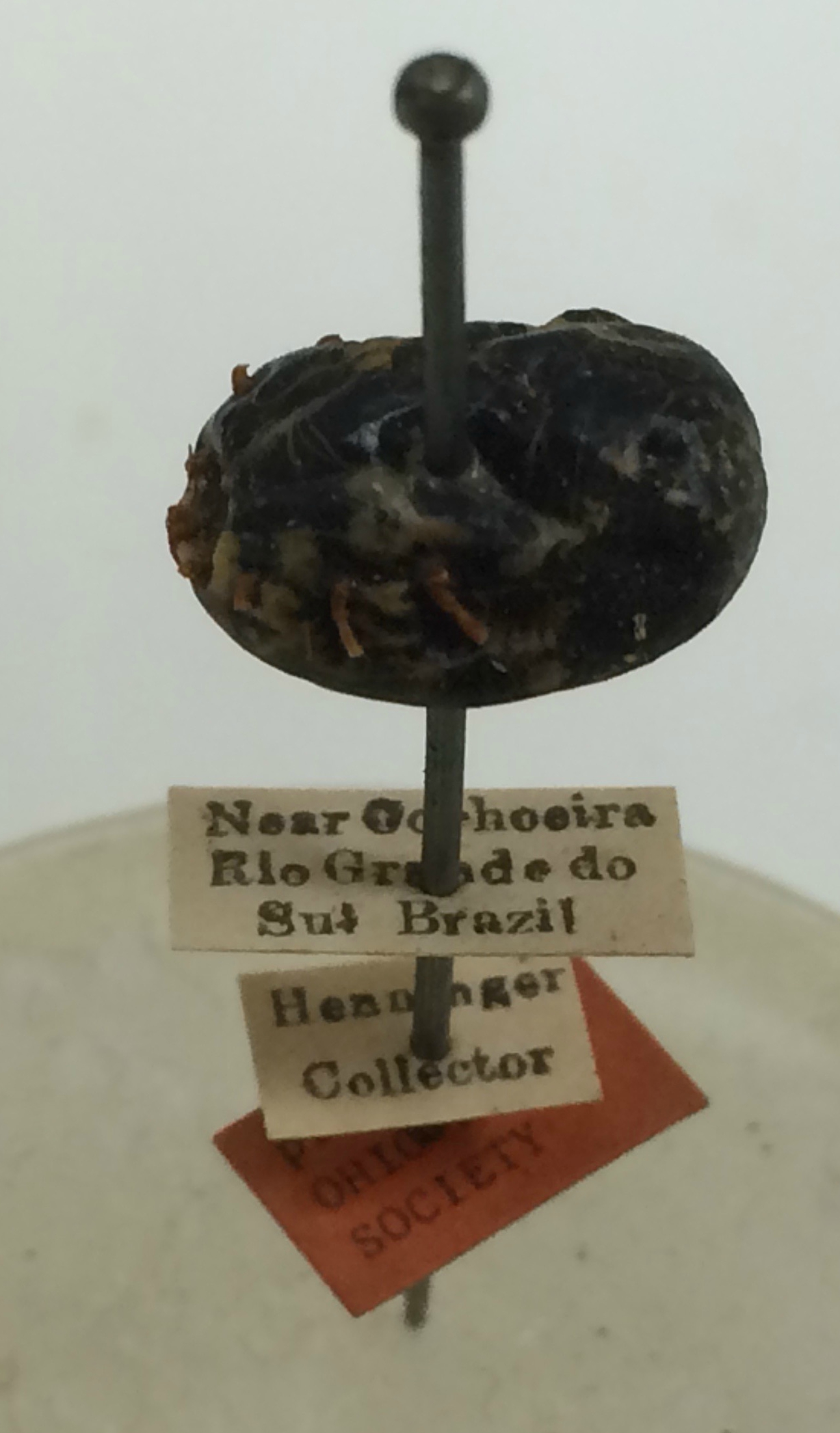
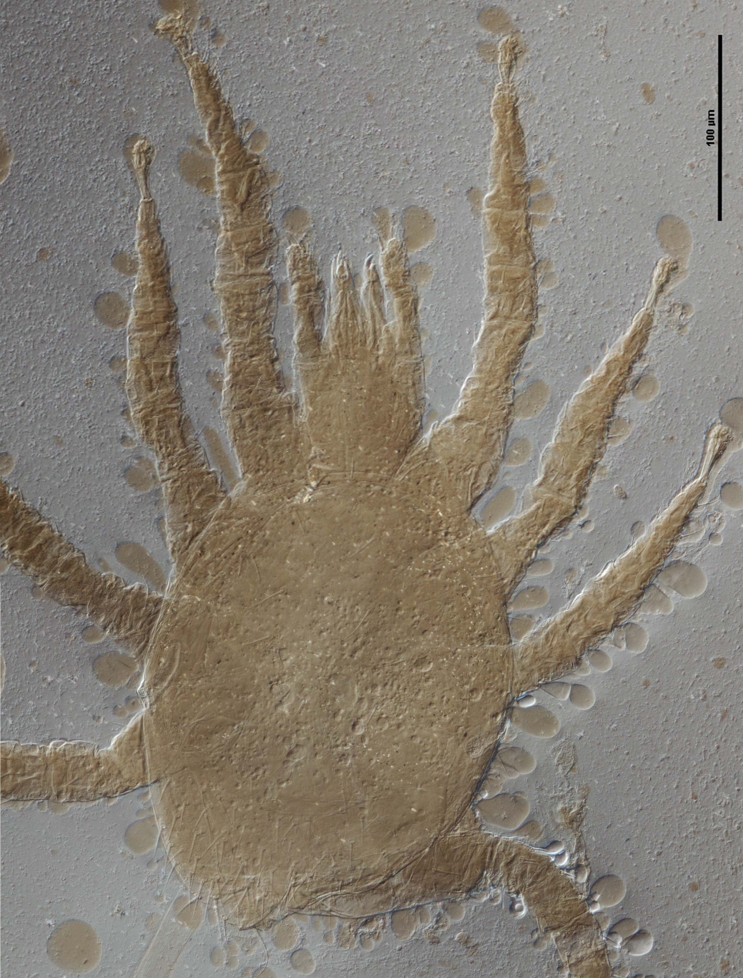
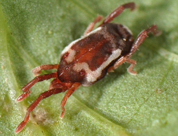
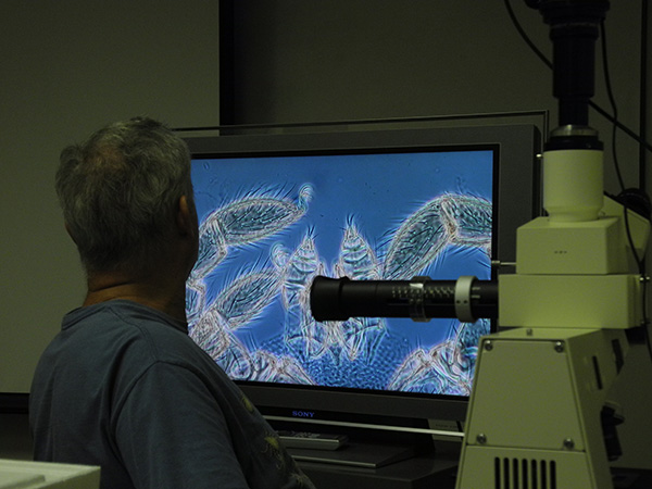
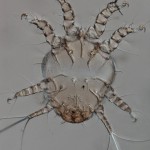
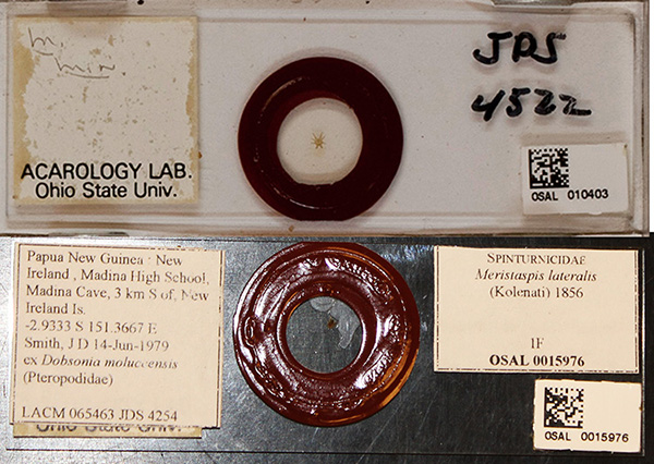

Yellow mite (Lorryia formosa)Acarology Laboratory.  Initiated by George W. Wharton in 1951, the Acarology collection is considered one of the best and most extensive insect and mite collections in North America. Over 150,000 identified and considerably more than one million unidentified specimens are included, preserved either in alcohol or on microscope slides. The geographic range is worldwide. The collection gets extensive use during the annual Acarology Summer Program, the foremost training workshop in systematic acarology in the world.

More information about the Acarology Lab can be found on their website. They also maintain the Acarology Summer Program website.


Borror Lab iconBorror Laboratory of Bioacoustics. The Borror lab is one of the leading collections of animal sound recordings in the United States. The Laboratory is named for Dr. Donald J. Borror, and entomologist and ornithologist who was a pioneer in the field of bioacoustics. He contributed many recordings including the first sound specimen in the archive, a recording of a blue jay in 1948. Today, the sound collection contains over 42,000 recordings, the majority of which are birds. Donald Borror also contributed many recordings of insects. Mammals, amphibians, reptiles, and even fish are part of the collection. The recordings are widely used for research, education, conservation, and public and commercial media.

Visit the Borror Lab website for more information and make sure to check their audio CDs.


Ohio Buckeye in bloom

Herbarium. The OSU Herbarium was founded in 1891 by Dr. William A. Kellerman,well-known botanical explorer of Central America, pioneer mycologist (that’s fungi!), and the University’s first professor of botany. It serves as a source of botanical data and as a base of operations for a wide variety of taxonomic, evolutionary, phytogeographical, and biochemical research programs; preserves specimens as vouchers to document present and past research studies or vegetation patters; serves as a reference point for the precise identification of plants, algae, protists, fungi and lichens; and serves the public by identifying plant specimens, providing morphological, systematic, and other information about plant species, and answering questions about plants, their properties and uses. The Herbarium currently holds over 550,000 specimens, including over 420 type specimens.

For more information about the Herbarium visit their website.


Molluscs icon

Molluscs. The Mollusc Division is really a collection of collections, containing over 1 million specimens in 140,000 lots. Over the years a number of private and institutional collections have been organized into the collection here today. The earliest large accession was that of Henry Moores (1812-1896) and was worldwide, both fossil and recent. Moores assembled one of the most diverse collections of labeled shells of that period. The University purchased this collection around 1890, added several private collections to it, and cataloged the material as part of the holdings of the first organization of the Ohio State University Museum in 1891. This collection and others were given to the Ohio State Museum on Campus in 1925, maintained and enlarged for nearly half a century, then returned to the administration of the University in 1970.

The Division of Molluscs has an interesting blog, Shell-fire and Clam-nation, and a website.


Tetrapod icon

Tetrapods. The Division of Tetrapods (amphibians, reptiles, birds, and mammals) is a repository of Ohio and North American species and some worldwide research expeditions. The collections were established shortly after the founding of The Ohio State University in 1870 and grew through the collecting efforts of OSU faculty. Specimens date as far back as 1837 and include many now-protected species as well as extinct species such as the Ivory-Billed Woodpecker, Carolina Parakeet, and Passenger Pigeon. The collection houses more than 170 amphibian, 200 reptile, almost 2,000 bird and 250 mammal species.

To learn more about the OSU Tetrapod collection, visit their website and their blog, Amphibians, Reptiles, Birds and Mammals.


Bowfin (Amia calva) skeleton

Fish. The Fish Division began with the collections of D. Albert Tuttle, OSU’s first zoologist. Officially recognized in 1895, the fish collection grew and moved from the Botany and Zoology Building to OSU’s Biological Station at Cedar Point, to the Ohio State Historical Society, to the Franz Theodore Stone Laboratory on Gilbraltar Island, to Sullivant Hall, and finally (whew!) to its current location as part of the Museum of Biological Diversity. The collection is primarily used as a resource for systematics research, laboratory teaching, and public education. It is also a resource for state and federal scientists who use it as a basis for comparative studies, document the geographic ranges of fish, and conduct ecological assessments and environmental impact statements.

Visit the Fish Division website for more information about their activities.

We hope you enjoy the blog and please send us your feedback!


About the AuthorDr. Norman F. Johnson is a Professor with appointments in the Department of Evolution, Ecology and Organismal Biology & the Department of Entomology at The Ohio State University. He is also the Director of the Triplehorn Insect Collection. Norman studies the systematics and evolution of parasitoid wasps in the family Platygastridae (Hymenoptera).