Ohio Dragonfly Survey – Spring Training 2018

Last Thursday, MaLisa Spring, state coordinator of the Ohio Dragonfly Survey, gave the introduction to a series of talks about dragonflies and damselflies, how to identify them (Bob Glotzhober), how to photograph them (Jim McCormac) and how to report them on iNaturalist (Jim Lemon).

The audience was captivated by stories such as dragonflies being ferocious hunters, some have even been reported to prey on hummingbirds (albeit rarely).

On the other hand, watching dragonflies glide over open water on a warm summer day can be very peaceful and give us appreciation for their beauty and flying ability.

Bob Glotzhober even speculated that the origin of the shape of the Valentine’s heart can be found in the mating ritual of some dragonflies. What do you think?

So how does one identify a dragonfly?

And how do you distinguish a dragonfly from a damselfly?

But be careful, size is not the only difference and may be deceiving: in the tropics some damselflies grow to 7 inches in length!

If you want to learn more about dragonflies, visit the Ohio Dragonfly Survey website or attend the Odonata conference in June 22-24 2018 in Findlay, Ohio.

https://u.osu.edu/ohioodonatasurvey/2017/11/08/save-the-date-for-odo-con-18-june-22-24-2018/

To identify dragons and damsels in the field, we recommend that you download the ODNR guide (booklet pub 320).

If you enjoy fishing, you may catch a dragonfly in its larval stage and the Atlas of the dragonfly Larvae may help you identify it.

As always, feel free to post any questions right here on our blog.

About the Author:  Angelika Nelson is the curator of the Borror Laboratory of Bioacoustics and the social media manager for the museum.

Follow us on Facebook.


Explaining Science – plant genomics

Brandon Sinn performing molecular lab work

Brandon Sinn performing molecular lab work

Brandon Sinn, PhD graduate from the OSU herbarium, now a postdoctoral fellow at West Virginia University, recently published a paper on molecular work he did to better understand the evolution of genomes in Asarum (Aristolochiaceae), commonly known as wild ginger. The work was done in collaboration with Dylan Sedmak, an OSU undergraduate student, Lawrence Kelly, Associate VP of Science, New York Botanical Garden and John Freudenstein, Professor and Chair of EEOB and Brandon’s PhD advisor.

We interviewed Brandon to get a better understanding of his research findings:

Brandon: “Evolutionary relationships in the flowering plant genus Asarum served as the focus of my dissertation research, and I continue to study the group.  In this particular project we studied six Asarum species, which each represent one of the six major evolutionary lineages within the genus.

Flowers of some Asarum species found in southern Appalachians

Flowers of some Asarum species found in the southern Appalachians

Asarum is a poorly-understood genus of approximately 115 species found in temperate forests across Asia and North America. Some Asarum species are common and widespread across the continents where they are found, while the majority have highly restricted ranges – for example, one species is known only from a single gorge in North Carolina and others are found in only a few counties in the southeastern United States.

During the course of sequencing DNA for my dissertation research, I realized that the genes of some Asarum species were not in the expected order. This departure from expectation was surprising, since the clade, or evolutionary neighborhood, that Asarum belongs to is very old and had been partially characterized as having slowly-evolving and highly conserved genomes. For example, the genome of another member of the same clade has been called a “fossil” genome. It was because of this unexpected observation that we decided to sequence complete genomes from one species from each of Asarum lineage. ”

This lead to the following research questions: Note: A plastome is the genome of a plastid, the organelle responsible for photosynthesis in plants.

1) Have the plastomes of all Asarum species been destabilized and their gene order rearranged?

2) Is the plastome of Saruma henryi (commonly called upright wild ginger), the closest relative of Asarum, of typical arrangement or is it more like that of Asarum?

3) Can we understand how the ordinarily highly conserved and stable plastomes become destabilized by comparing the plastomes of many Asarum species to that of Saruma henryi?

Saruma henryi, a flowering plant in the family Aristolochiaceae, endemic to China

What should we know to understand this research?

Brandon: “Each plant cell contains at least one copy of three distinct genomes. It is easy to imagine that each cell has a copy of the plant’s genome, but many people forget that two types of plant organelles, mitochondria and plastids, also have their own genomes. Plastids, from which chloroplasts develop, have a very small genome that is relatively easy to completely sequence and the sequence of more than 2,000 are publicly available today. The sequencing of thousands of plastomes has resulted in several general trends: 1) plastomes change more slowly than the plant’s own genome; 2) the plastome is made up of three functional regions, the small single copy, large single copy, and inverted repeat regions; 3) the physical order of genes is highly conserved across even distantly related species; 4) there is very strong selective pressure on the preservation of photosynthesis, which most likely constrains the evolution of plastomes in green plants. Our knowledge of the typical layout of the plastid genome, or plastome, has long been relied upon to sequence DNA in order to study plant evolutionary relationships. Traditional DNA sequencing techniques require prior knowledge of the order of genes or regions of a genome. If this order is not as predicted, then the DNA sequencing will fail.”

What method did you use to study your research question?

Brandon: “For this study, we sequenced entire plastomes from six Asarum species and that of Saruma henryi, the closest relative of Asarum. Since traditional DNA sequencing is not useful in destabilized and dynamically rearranged genomes, and we wanted to sequence entire plastomes that we hypothesized were rearranged, we needed to use a technology called massively parallel sequencing. A major advantage of massively parallel sequencing is that a researcher can extract DNA from a tissue, break the DNA into short pieces, and simultaneously sequence all of these fragments without prior knowledge of their physical relationship to one another. The resulting millions of DNA sequences are then assembled, much like a puzzle, using specialized software. The assembled  plastomes can then be compared.”

Brandon explains one of the key figures in his manuscript:

A cruciform DNA structure that has likely destabilized a region of the plastome in Asarum species. Structure courtesy of Eric Knox.

A cruciform DNA structure that has likely destabilized a region of the plastome in Asarum species. The end of the ndhF gene is shown in red. Structure courtesy of Eric Knox.

DNA is made of only four chemicals (which we abbreviate as the letters A, T, C and G) and is not entirely unlike a spiral staircase, where each handrail is a string of these letters. Holding this structure together are bonds that form between certain letters – A-T and G-C. We call these letters nucleotides. Sometimes the nucleotides making up DNA cause the molecule to form complex shapes, such as the cruciform structure shown here. Cruciform, or cross shaped, DNA structures form when the same nucleotides are repeated very close to one another, which is depicted in the vertical “stems”.

plastomes

Cruciform DNA structures can be difficult for the molecular machinery in cells to work with. For example, sometimes molecules that interact with DNA get stuck on the stems, and these structures compromise the integrity of the DNA molecule. When these structures break, which you can imagine by separating the red and black halves of DNA for Saruma henryi, the cell tries to put them back together. But, repairing DNA does not always work perfectly. The results of our research suggest that faulty repairs made to this DNA structure throughout the plastomes of Asarum species have resulted in varying degrees of DNA duplication. Notice that the ndhF gene (shown in red) is typically at one end of the small single copy region, as shown on the Saruma henryi plastome. In Asarum, this gene often has a long stretch of nucleotides that can be “pasted before or after it. In other Asarum plastomes, such as Asarum canadense, we find that all of the small single copy region has been duplicated. The duplication of the formerly single copy region is most likely due to faulty repair of the cruciform DNA structure, where identical strings of nucleotides close to one another led to bonding of two identical DNA regions (as seen in the Asarum canadense cruciform structure).”

Why is this research important?

Brandon: “When you learn about DNA in high school science classes, everything sounds very concrete and well understood, but even gene function in humans is not exhaustively understood. Our basic knowledge about how genes and genomes evolve is in a constant state of improvement. This knowledge is necessary for future breakthroughs in genome engineering, evolutionary and conservation biology, and improving genome stability.  Just as it is important to understand biodiversity at the level of species, it is equally important to understand genomic diversity – the content and structure of genomes, in order to understand how mutations in particular regions of genomes can lead to genome-scale changes over deep time and how these changes affect evolutionary lineages.”

What should you take away from these findings?

1) Just because a species is a member of a very old evolutionary lineage, we should not expect that it is a living fossil and that its genome has changed little.

2) A plastome can function even when gene order is changed and more than half of its genes are present more than once.

3) Small, likely randomly generated repetitive motifs in DNA sequence that is not part of a gene can decrease genome stability, and lead to genome rearrangement and gene duplication.

*******************************************************

Wow, we are now certainly asking questions and getting answers with new techniques that we could not have imagined decades ago. If you want to follow Brandon’s further research, click here.

About the Authors: Brandon Sinn photoBrandon Sinn earned his Ph.D. in 2015 from the Department of Evolution, Ecology and Organismal Biology, where he was a member of the Freudenstein Lab in the Museum of Biological Diversity. Brandon has held a postdoctoral research position at the Pfizer Plant Research Laboratory of the New York Botanical Garden, where he worked on the Planteome Project. He is currently a postdoctoral fellow in the Department of Biology of West Virginia University where he studies orchid genome evolution as a member of the Barrett Lab.

Angelika Nelson is the curator of the Borror Laboratory of Bioacoustics and the social media manager for the museum.

Follow us also on Facebook.


Reference:
Sinn, B. T., Sedmak, D. D., Kelly, L. M., & Freudenstein, J. V. (2018). Total duplication of the small single copy region in the angiosperm plastome: Rearrangement and inverted repeat instability in AsarumAmerican journal of botany105(1), 71-84.

Interview with the coordinator of the Ohio Dragonfly Survey

MaLisa SpringMaLisa Spring, State Coordinator for the Ohio Dragonfly Survey, sat down with us to talk about the Ohio Dragonfly Survey and its focus – dragonflies and damselflies!

Hilary: “Tell us about yourself!”

MaLisa: “I completed a Bachelor’s in Biology with a minor in Spanish at Marietta College, Ohio. While I was there I did a couple of research projects related to insects and completed my senior thesis on bee diversity. It was then that I decided that I wanted to go to grad school, and I ended up attending OSU, where I received my Master’s degree in Entomology.

I worked with native pollinators for my undergraduate, but I also did a small project on lady beetle diversity and completed an internship on insect diversity. Overall, I’m just broadly interested in entomology and so when there was a mention of a dragonfly survey coming up and that they were interested in hiring someone for the survey, I was all for it – it sounded great! I then landed the job as the state coordinator for the Ohio Dragonfly Survey in May of 2017.

Hilary: “What is the Ohio Dragonfly Survey and what is its goal?”

MaLisa: “The Ohio Dragonfly Survey is a citizen science project with the goal of getting people outside, to notice dragonflies and damselflies, and to submit their observations to the survey via iNaturalist.org. Our goal right now is to figure out where species are throughout the state and to document the status of the threatened and endangered species. Ohio has 167 species of dragonflies and damselflies and we have 23 species that are state-listed as either endangered, threatened, or a species of concern, but there could be several more species added to that list. Additionally, the Hines Emerald (Somatochlora hineana) is the only federally endangered species in our state and it hasn’t been seen since at least 1989, so it might be extirpated (locally extinct, but surviving elsewhere).”

Double-striped bluet (pond damselfly)

Double-striped bluet

Hilary: “What are some of the greatest threats to dragonflies?”

MaLisa: “There are many threats to dragonflies and damselflies. Habitat loss and degradation are two of the biggest ones. Compared to the species richness between now and say the 1950s, there’s a huge difference. For example, Ohio used to have the Great Black Swamp, a several county-large swamp up in northwestern Ohio, but it was drained and turned into agricultural fields. Reducing hectares of wetlands into ditches created a vastly different habitat for the dragonflies and damselflies of this region, resulting in a significant change in the number of species that used to live here.

Other challenges are pesticides and especially herbicide run off.  Herbicides affect plants and certain species of dragonflies and damselflies lay their eggs inside of specific plants. If those plants aren’t there for them to lay eggs in, then the species cannot survive. They also need plants at the edge of water, so if people are mowing down plants at the edge of ponds or other ideal aquatic habitats, then the dragonflies and damselflies don’t have anywhere to emerge after their larvae stage to spread their wings to dry, and so they die.”

Hilary: “What is the life cycle of a dragonfly or damselfly?”

MaLisa: “Immature dragonflies and damselflies, also referred to as water nymphs (naiads) or larvae, reside in the water or aquatic systems, where they can live anywhere between 3 weeks to several years (it depends on the species). Some species are migratory, so they migrate to and from Ohio, but others overwinter in Ohio as larvae in the water systems and then emerge in either the spring or summer (again, it depends on the species) as adults, where their lifespans can range from two weeks to up to a couple of months.

Hilary: “How can you tell a dragonfly and a damselfly apart?”

MaLisa: “Dragonflies and damselflies are in the order odonata and are divided into two separate groups, Anisoptera and Zygotpera. You can differentiate the two based on the wing positioning for the most part, but not always. Dragonflies tend to hold their wings out like a biplane, whereas damselflies tend to hold their wings behind their back like a sailboat.

Another way to differentiate them is based on their size. Most dragonflies are larger than damselflies. But again, there’s an exception to the rule with the body of some damselflies being longer than that of some dragonflies. If you want to get into the nitty gritty to tell them apart, you can look at their eyes to see how separated they are from each other. All damselflies have their eyes well separated on their head, whereas most dragonflies (excluding the Clubtails) have their eyes at least touching.”

Hilary: “For the public to get involved in the survey, do they need to collect specimens?”

MaLisa: “For the most part, we’ve learned that the public doesn’t want to collect specimens, which is okay as photographing them instead is perfectly acceptable. However, if they are interested in collecting specimens we ask that they look on our webpage and follow our collection protocols.”

Hilary: “Will you be at the Open House for the Museum of Biological Diversity on April 7th?”

MBD Open House April 7th, 2018

MaLisa: “I will definitely be here for the Open House event, if anyone wants to ask me any dragonfly questions!”

To learn more about the Ohio Dragonfly Survey, access this link: http://u.osu.edu/ohioodonatasurvey/

Contributing to the survey is as easy as taking photos of dragonflies and damselflies and submitting them to iNaturalist.org! Learn more here.

Want to learn more about dragonflies and damselflies? Check out this page for upcoming talks, presentations, and community events. Or download a guide to dragonflies and damselflies of Ohio from the Ohio Division Wildlife.

Hilary HirtleAbout the Author: Hilary Hirtle is the Faculty Affairs Coordinator at the OSU Department of Family Medicine; her interest in natural history brings her to the museum to interview faculty and staff and use her creative writing skills to report about her experiences.

Staff spotlight – Scott Glasmeyer

We met up with Scott Glassmeyer, a student research assistant in the Fish Division, to get an inside view on his role in the Museum of Biological Diversity.

Scott Glassmeyer holding a Rock Bass (fish)Hilary: “What is your major?”

Scott: “My major is Forestry, Fisheries, and Wildlife, with a specialization in Fisheries and Aquatic Science. I’d always loved fish since I was a kid and before I got into this program, I didn’t know that you could go to college to study fish, or do anything relevant with fish in a job, besides working to commercially collect fish. So, I did research to see if there were any higher education programs that involved learning about fish and aquatics and I found that Ohio State had this program.”

Hilary: “How long have you been a student research assistant in the Fish Division?”

Scott: “Since spring of 2016, but I started as a volunteer in January of 2016 where my primary role was to take older jars containing fish specimens and place them in new ethanol, to better preserve the fish.”

several fish in ethanol in glass jar

Fish in ethanol

Hilary:  “What is the mission of the Fish Divsion?”

Scott: “To preserve historical records of species of fish for future reference and overall long-term data collection and education. It’s a way to validate that this species of fish was recorded in a particular area and a specific species was recorded in general, as fish get misidentified a lot. So it improves a lot of accuracy regarding records.”

Hilary: “What fish are housed here?”

Scott: “Mostly Ohio fish, but we have some from the entire 50 states as well. There are also some fish species from other countries, some saltwater fish, and some aquarium fish here as well.”

Hilary: “Are the specimens here largely donated?”

Scott: “A lot of the specimens are collected through the museum, as well as the Ohio EPA. The Ohio EPA has a division that monitors streams and stream quality statewide and they will collect fish in the process and send them to us.”
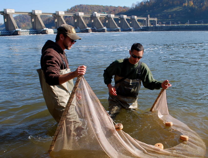
collecting fish with seine nets

Staff collecting fish with seine nets


Hilary: “How are the fish preserved?”

Scott: “The way the preservation process works is that you put the fish specimens in formaldehyde for a certain amount of time, then you place them in water for about a day, before you start adding the ethanol bit by bit, as you slowly add larger amounts of ethanol to build up the tolerance – and that’s what they stay in. It takes up to a week and a half to two weeks to put them in this preserved state.”

Hilary: “Why is it important to study these fish?”

Scott: “It’s really important to study these fish because it helps you not only understand the water quality of their habitat, but also the intrinsic value of their ecosystems. For example, if you have a stream that’s just concrete because it was filled in, this could possibly only allow for about 5 species of fish to live there, whereas before, when the stream had natural morphological features and geological shapes, there were a lot more species of fish living within in this habitat.”

“A good example of this is from about 6 or 7 years ago, when the 5th Avenue Dam along the Olentangy River near campus was removed. Trees, plants, and wetlands were added along the bank and this natural state contributed to the value of the stream, not just for people, but for the fish as well, as this improved quality increased the level of biodiversity within in and around the river.”
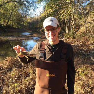
Scott Glassmeyer holding Giant bottlebrush crayfish

Scott Glassmeyer holding a Giant bottlebrush crayfish

Hilary: “What’s your favorite part about working in the Fish Division?”

Scott: “I love going outside, putting waders on, getting in the stream and finding fish. You can read all you want about how healthy a stream is, but when you go out there and you see the biodiversity in the water as you collect data, you can tell just how healthy the water is and it’s wonderful.”

“I also really like the people who work here with me. Everyone’s very patient here and they take the time to help you out as your learning, which is really nice as learning to identify fish for the first time involves a learning curve.”

Hilary: “What is a project that you’re working on now?”

Scott: “I’ve been editing photographs of fish taken by Brian (my colleague who is the Sampling Coordinator in the Fish Division) and getting them ready to be put into the field guide version of the Fishes of Ohio.”

book cover Fishes of Ohio by Milton B Trautman

“The Fishes of Ohio was a guide written in the ‘50s, by Trautman, and then it was revised in the ‘80s by Trautman, and so what we’re working on now would be the next revision. There’s around 190 species or so of fish in Ohio, including invasive species and extinct species, so we’ve been photographing each species listed in the field guide, oftentimes with more than one picture, as you’re taking pictures of what you use to identify them. For example, for some of the sucker species of fish, you have to show the mouth, as that helps with identification. So with these species, there’s some photographs detailing the mouth from underneath, and there’s some side photographs, so that you can see the shape of the head and the mouth from the side for identification.”


Hilary: “Do you photograph the fish in their habitat?”

Scott: “It depends. There was one species of fish where we went out during their spawning season and had the tank set up to photograph them. We caught them, put them in the tank, and took a picture quickly, as they can lose their colors pretty fast. If a fish we find doesn’t have a particular color, we take them, put them in a cooler with an aerator, and take them away from location to photograph them. It’s a time consuming process, with the drive to the specimen’s location, the set-up, hours of wading for fish, and then the tear-down of equipment and the drive back from the site, so taking them away to photograph them can be easier than doing it onsite.

Hilary: “You said that fish lose their colors – what does that mean?”

Scott: “Fish have pigments in their skin, underneath their scales. There’s a lot of colorful fish in Ohio, like darters and minnows, that will have breeding colors and so, during certain times of the year and certain times of the day (or even after they eat) they’ll get a lot of pigment and colors in them. And even if they’re not a colorful fish, their colors can change. For example, you can take a large mouth bass that has some pattern to it and put it into a bucket that’s really light and pull the fish out ten minutes later, and the fish will look really pale. But if you put it in a dark cooler, the fish is going to remain dark and have more color. The stress levels will impact them.”

Hilary: “Do you have a favorite fish species?”

Scott: “This question’s hard. So, my answer changes every month when I discover a new fish, but currently my favorite fish is the Common Dolphin Fish, or the Mahi-mahi. There’s a reason why I like it: So, over 50% of its diet is flying fish, and that’s pretty cool to me. Also, its maximum life span is five years. A marlin or a swordfish can live to be about 27 years of age, and a medium sized Ohio fish species can live to about 15 years. However, the Dolphin Fish lives such a short span of time compared to these fish, yet it grows extremely quickly, as they get up to 36 pounds in 8 months. And it’s really fast too, swimming speeds up to 50 miles an hour.”

Hilary: “With all of your experience and studies, what do you hope to do in the future?”

Scott: “I’d love to work as a fisheries biologist, working for the environment. It’s challenging to get in those types of roles, as they’re very competitive, but I’m going to try.”


Hilary HirtleAbout the Author: Hilary Hirtle is the Faculty Affairs Coordinator at the OSU Department of Family Medicine; her interest in natural history brings her to the museum to interview faculty and staff and use her creative writing skills to report about her experiences.

A Snapshot of Ohio Lichen Diversity 125 Years Ago

The Kellerman Displays for the 1903 Chicago Exposition

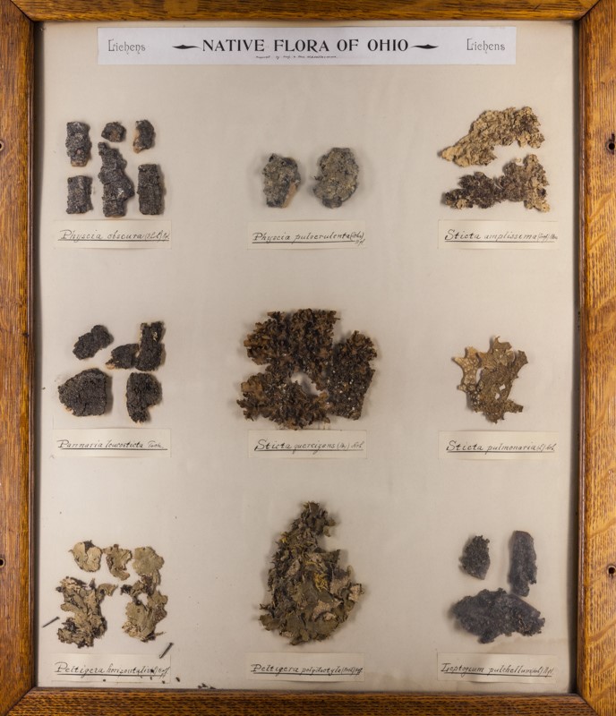
Most of the specimens at the Ohio State University Herbarium (OS) are tucked neatly into cabinets, not on display. But adorning one long wall are what at first glance look like pictures. Artfully arranged, with wood frames and a glass front, a close look reveals they are not paintings but are in fact real, once-living, plants and fungi.

Framed specimens at The Ohio State University Herbarium

The displays are quite pretty and they’re obviously rather old, but I never stopped to consider just how old they are, or how they came to be. A modern interpretive sign explains that they, along with four larger, more intricate panels of Ohio trees, were assembled for display at the World’s Columbian Exposition, a big world’s fair held in Chicago for six months in mid-1893.

write-up by Ronald L Stuckey about Kellerman's Columbian exposition mounts

Write-up by Ronald L Stuckey about Kellerman’s Columbian exposition mounts

At the top of each 18 x 22-inch panel is a printed heading “Flora of Ohio,” and beneath that, in ornate old-style penmanship, are the words “Prepared by Professor and Mrs. W. A. Kellerman.” William A. Kellerman was remarkably energetic and wide-ranging in his botanical interests. Making these panels was an appropriate hobby for a person whose life revolved around plants and fungi. An Ohio native born in 1840, he attended Cornell University for undergraduate studies and later received his Ph.D. from the University of Zurich, Switzerland. He taught at schools in several nearby US states before returning home to become OSU’s first botany professor and Chairman of the Department of Botany when it was formed in 1891. That same year, he established the Herbarium in a building aptly named “Botany Hall” that unfortunately no longer exists on OSU’s oval. Since then the Herbarium has moved twice, first to the also aptly named “Botany and Zoology” building (now Jennings Hall) and then to its present location as part of the Museum of Biological Diversity on West Campus (1315 Kinnear Rd.). While his principal research interest was rust fungus diseases of crops, Kellerman’s numerous works on the flora of the regions where he lived reveal an extraordinary breadth of knowledge. He wrote a guide intended principally for use by teachers entitled “Spring Flora of Ohio” (1895) and co-authored, beginning in 1894 and subsequently updated several times, “A catalogue of Ohio Plants.” Sadly, while Kellerman was on a research trip to study fungi in Guatemala, he contracted a fever (most likely malaria) from which he died in 1907.

Photo of WA Kellerman in the Journal of Mycology

Photo of W.A. Kellerman in the Journal of Mycology

The panels are an interesting snapshot of the flora of Ohio. While aesthetics and enthusiasm for particular plants may have played a major role in their selection by the Kellermans, the panels were indeed portrayed to fairgoers as indigenous representatives of our flora. As there have been substantial changes in the composition of our vegetation, especially for such pollution and disturbance-sensitive organisms as lichens, they arouse curiosity about the past versus present status of these organisms.

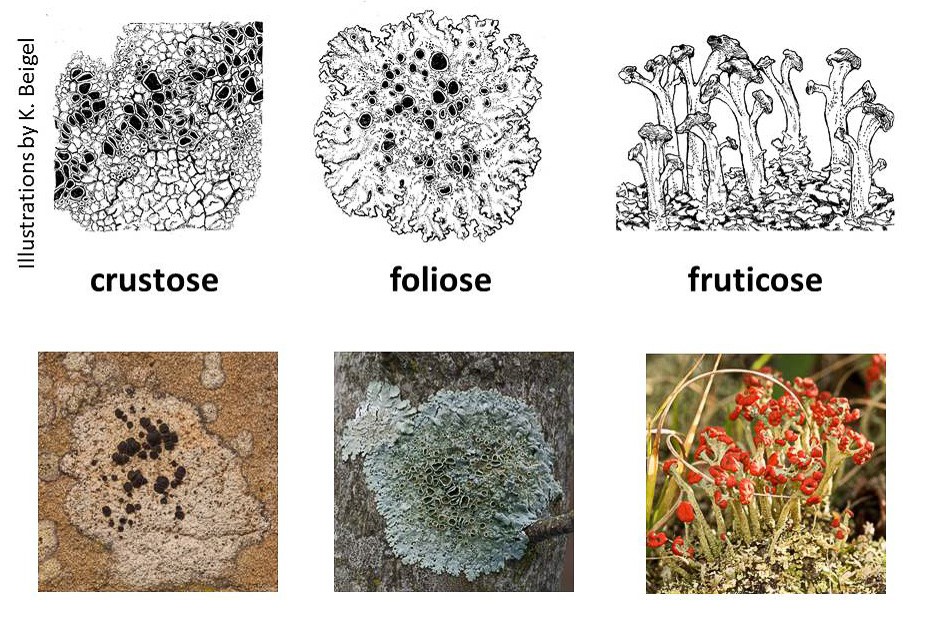
Lichens are dual organisms consisting of fungus plus alga. The algae are single-celled photosynthetic organisms. The fungus, which constitutes most of the body of a lichen, provides a home for the algae, usually in a layer just beneath the surface. Most lichens fall into one of three growth-form categories: (1) usually small “crustose” lichens that are tightly attached to the substrate and so don’t have a discernable lower surface; (2) small to medium-sized “foliose” lichens that are flattened and can usually be separated from the substrate, and (3) “fruticose” lichens that have a bushy shape, either standing upright from the surface they are growing on, or dangling off a tree branch or trunk. Most of the lichens in the panels are foliose species.

Illustration of three growth-form categories of lichens

Three growth-form categories of lichens

There doesn’t seem to be a strict organization scheme for the lichen panels; they’re not in alphabetical or taxonomic order, except that one panel consists mostly of crustose species, while the few fruticose ones represented are grouped together, sharing space with some foliose ones. I suspect that the paucity of fruticose types is attributable to the display method only being suitable for flat or readily flattened specimens.

Each panel includes 9 specimens, with handwritten labels. The classification of lichens has undergone substantial change in the past century and a quarter, hence many of the names written by the Kellermans are not in use today. Fortunately, an on-line database called “Consortium of North American Lichen Herbaria” lists specimen records for lichens residing in collections spanning the continent, and the site lists all the names by which a species has been known in the past.

The present distribution of lichens in Ohio is well described in The Macrolichens of Ohio by Ray E. Showman and Don G. Flenniken, published in 2004 by the Ohio Biological Survey, and distribution maps presented on the web site of the Ohio Moss and Lichen Association. The status of the lichens more broadly is set forth in a monumental book, Lichens of North America by Irwin M. Brodo, Sylvia D. Sharnoff and Stephen Sharnoff, published in 2001 by Yale University Press, along with an updated companion volume by Brodo published in 2016 by the Canadian Museum of Nature, Keys to Lichens of North America: Revised and Expanded.

One panel caught my eye. This is a group of mostly rather large foliose lichens, including several “lungworts,” members of the Lobaria –robust broad-lobed species found on bark.

Display of a group of mostly rather large foliose lichens

A group of mostly rather large foliose lichens

Among the most easily recognized of all lichens, lung lichen, Lobaria pulmonaria, was once widely distributed across Ohio, but no more. All but one of the 14 county records for lungwort are pre-1945, with the other one record being sometime between 1945 and 1965. Extensive searching has failed to find lung lichen today.

Why is it lung lichen gone from Ohio? It’s probably due to a multiplicity of factors that prevailed during the late 19th, and early 20th centuries: air pollution and disturbance of old-growth forests. Now that conditions are better for it to grow, perhaps a lack of propagules is keeping it from reestablishing itself. While eventually a warbler or vireo might fly in from some north woods with a little piece of lungwort on its foot, this might be a good candidate for a deliberate reintroduction.

Photo of lungwort growing on a tree in Maine

Lungwort growing on a tree in Maine

This is what lungwort looks like, growing on a tree in Maine. It’s a beautiful lichen and that just might still be growing in in a bottomland forest somewhere in Ohio, or it might soon return. Keep an eye out for it the next time you go hiking!

About the Author: Bob Klips is Associate Professor Emeritus in the department of EEOBiology at The Ohio State University. He currently assists with moss and lichen databasing in the OSU herbarium. His research focuses on bryophyte ecology.

Explaining Science – Gene flow among song dialects

Today Kandace Glanville, an OSU Forestry Fisheries & Wildlife major and student assistant in the Borror Laboratory of Bioacoustics, talks with Angelika Nelson, Curator of the Borror Lab, about a recent research publication in the journal Ethology. The study is entitled “High levels of gene flow among song dialect populations of the Puget Sound white-crowned sparrow”.

Find out why we studied the White-crowned Sparrow Zonotrichia leucophrys pugetensis to investigate gene flow among song dialects:

The research aimed to investigate a correlation between behavioral and genetic differentiation:

Our research built on knowledge from previous studies and used samples that were collected previously:

We found gene flow among bird populations that differ in song dialects; this may demonstrate dispersal of young birds across dialect borders:

Our findings are consistent with most studies to date of song and population structure within songbirds. The processes of song learning and dispersal mean that vocalizations are free to vary independently of patterns of divergence in neutral genetic markers.

Reference:
Poesel, Angelika, Anthony C. Fries, Lisa Miller, H. Lisle Gibbs, Jill A. Soha, and Douglas A. Nelson. “High levels of gene flow among song dialect populations of the Puget Sound white‐crowned sparrow.” Ethology 123, no. 9 (2017): 581-592.


About the Author: Angelika Nelson is the curator of the Borror Laboratory of Bioacoustics and the social media manager for the Museum of Biodiversity.

Explaining Science – vermiform mites

You have heard of mites – minute arachnids that have four pairs of legs when adult, are related to the ticks and live in the soil, though some are parasitic on plants or animals. But what are vermiform mites? Maybe you have heard of vermi-compost, a composting technique that uses worms (like your earthworm in the garden) to decompose organic matter. So vermiform mites are mites with a body shape like a worm:

worm-shaped nematalycid Osperalycus

Why are they shaped like a worm, you may ask – To find out more I interviewed Samuel Bolton, former PhD student in the acarology collection at our museum, now Curator of Mites at the Florida State Collection of Arthropods. Sam’s main research interest is in mites that live on plants and in the soil, especially Endeostigmata, a very ancient group of mites that dates back around 400 million years, before there were any trees or forests. Sam’s PhD research with Dr. Hans Klompen here at OSU, was focused on a small family (only five described species) of worm-like mites, called Nematalycidae.

side note: You may have heard of Sam’s research in 2014 when he discovered a new species of mite, not in a far-away country, but across the road from his work place in the museum.

When Sam started his research it was not clear where these worm-like mites in the family Nematalycidae belong in the tree of life. To find out Sam studied several morphological characters of Nematalycidae and other mites. He focused in particular on the mouth-parts of this group. As he learned more about the mouth-parts of this family, he found evidence that they are closely related to another lineage of worm-like mites, the gall mites (Eriophyoidea). Eriophyoidea have a sheath that wraps up a large bundle of stylets. They use these stylets to pierce plant cells, inject saliva into them and suck cell sap.
Although Nematalycidae don’t have stylets, one genus has a very rudimentary type of sheath that extends around part of the pincer-like structures that have been modified into stylets in Eriophyoidea.

So what did Sam and his co-authors discover?

“.. Not only are gall mites the closest related group to Nematalycidae, but the results of our phylogenetic analysis places them within Nematalycidae. This suggests that gall mites are an unusual group of nematalycids that have adapted to feeding and living on plants. Gall mites use their worm-like body in a completely different way from Nematalycidae, which live in deep soil. But both lineages appear to use their worm-like bodies to move around in confined spaces: gall mites can live in the confined spaces in galls, under the epidermis (skin), and in between densely packed trichomes on the surface of leaves;  Nematalycidae live in the tight spaces between the densely packed mineral particles deep in the soil.”

This research potentially increases the size of Sam’s family of expertise, Nematalycidae, from 5 species to 5,000 species. We have yet to confirm this discovery, but it is highly likely that gall mites are closely related to Nematalycidae, even if they are not descended from Nematalycidae. This is interesting because it shows that the worm-like body form evolved less frequently than we thought. This discovery also provides an interesting clue about how gall mites may have originated to become parasites. They may have started out in deep soil as highly elongated mites. When they began feeding on plants, they may have used their worm-shaped bodies to live underneath the epidermis of plants. As they diversified, many of them became shorter and more compact in body shape.

I wish I could tell you now to go out and look for these oddly shaped mites yourself, but you really need a microscope. Eriophyoid mites are minute, averaging 100 to 500 μm in length. For your reference, an average human hair has a diameter of 100 microns.

eriophyoid Aceria anthocoptes

Reference:

Bolton, S. J., Chetverikov, P. E., & Klompen, H. (2017). Morphological support for a clade comprising two vermiform mite lineages: Eriophyoidea (Acariformes) and Nematalycidae (Acariformes). Systematic and Applied Acarology, 22(8), 1096-1131.


About the Authors: Angelika Nelson, curator of the Borror Laboratory of Bioacoustics, interviewed Samuel Bolton, former PhD graduate student in the OSU Acarology lab, now Curator of Mites at the Florida State Collection of Arthropods, in the Florida Department of Agriculture and Consumer Services’ Division of Plant Industry.


Bat sounds

Bats are social mammals that use a repertoire of vocalizations to communicate with each other and to move around in the environment.

To detect obstacles and prey in their environment, bats emit a series of ultrasounds, very high-pitched sounds above 20,000 Hz, beyond our range of hearing. As a bat flies and calls, it listens to the returning echoes of its calls to build up a sonic image of its surroundings. Bats can tell how far away something is by how long it takes the sounds to return to them, how big the target is based on the strength of the returning signal, and what shape the target has based on the spectral pattern of the returning sound waves. We call this process echolocation.

Individual bat species echolocate within specific frequency ranges that suit their environment and prey types. This means that we can train ourselves to identify many bats by listening to their calls with bat detectors.

Let’s LISTEN to recordings of the little brown bat (Myotis lucifugus) and the big brown bat (Eptesicus fuscus) for comparison. – But how can we listen, if we cannot hear their calls? Let’s use a trick: When we slow down the recordings by a factor of 10, the calls are transposed to 10 times lower pitch and become audible to us.

Note: To make the sounds visible in sonograms we plotted frequency in thousands of cycles per second (kilohertz, kHz) on the vertical axis versus time in seconds on the horizontal axis. The varying intensity of colors ranging from dark blue (low intensity or quiet) to red (high intensity or loud) indicates the amplitude or loudness of each call. Amplitude is also shown in the top part of each figure with larger waves representing louder calls.

Little brown bat: Calls last from less than one millisecond (ms) to about 5 ms and sweep from 80 to 40 kHz, with most of their energy at 45 kHz.

sonogram of little brown bat Myotis lucifugus calls

Call series of a little brown bat Myotis lucifugus


Big brown bat: Calls last several milliseconds and sweep from about 65 to 20 kHz, and are thus lower pitched than calls of little brown bats.

bigsonogram of brown bat Eptesicus fuscus echolocating calls

Call series of a big brown bat Eptesicus fuscus


The above call series were recorded when the bat is generally surveying its environment, but what happens when it actually detects prey? Listen to this feeding buzz of a little brown bat:

sonogram of feeding calls of little brown bat

Feeding calls of a little brown bat Myotis lucifugus


When closing in on prey, a bat may emit 200 calls per second.

What might sound to us like the bat is getting excited – don’t you talk faster when you are excited about telling something? – this rapid series of calls actually helps the bat to pin-point the exact location of its prey, then it swoops in, and GULP – dinner is served, or not!


We hope you enjoyed listening to these bat sounds; if you have any questions please contact Angelika Nelson.794@osu.edu, curator of the animal sound archive at The Ohio State University.

The Ohio State University - logo


All recordings are archived with the Borror Laboratory of Bioacoustics (BLB.OSU.EDU) at The Ohio State University.

A gull look-alike

Another seabird species that I found to breed in Ireland is the Northern Fulmar Fulmarus glacialis. In a fleeting glimpse this bird may look like a gull but a closer look quickly reveals that is a close relative of albatrosses and shearwaters, the tubenoses Procellariiformes.

Can you see how this group of birds, the tubnoses, got its name? Doesn’t it look like they have a tube on top of their bill? This tubular nasal passage is used for olfaction. Yes, some birds do have the ability to smell. Especially seabirds use this sense to locate flocks of krill, shrimp-like animals that feed on single-celled marine plants (phytoplankton) right below the ocean’s surface. Breaking up phytoplankton cells releases a chemical called dimethylsulfide that concentrates in the air above areas where phytoplankton and thus krill are abundant. Researchers suspect that seabirds may smell their prey.

An acute sense of smell may also aid these birds to locate their nest within a breeding colony – you may recall the dense breeding conditions on the coastal cliffs from Monday’s post.

Furthermore, at the base of their bill these true seabirds have a gland that helps them excrete excess salt as they drink seawater. These birds and their relatives often spend long times out over the ocean without any land in sight. Thus they depend on drinking seawater.

So what do Northern Fulmars sound like? They are especially vocal when they return to their partner on the nest, they engage in an often minutes-lasting greeting ceremony. Listen to this pair recorded by Gabriel Leite in Clare county, Ireland (XC372370):

The unique morphological characteristics make these birds well adapted to their preferred environment of the northern oceans. They are among the longest-lived birds known, researchers estimate an average lifespan of 32 years for the Northern Fulmar.

About the Author: Angelika Nelson is the curator of the Borror Laboratory of Bioacoustics and currently teaches at the Audubon summer camp on Hog Island, ME.


Playing the role of a bee

Mid-spring through mid-summer is a good time to see our native orchids in flower here in Ohio.  One of the showiest groups is the Lady’s Slippers, which have a distinctive pouch-shaped lip.  We have four species of Lady’s Slippers (Cypripedium) in Ohio and one of the more frequent ones is the Yellow Lady’s Slipper (C. parviflorum).  There are two varieties of this species – Large and Small.  The Large (var. pubescens) tends to be a plant of rich woods in more upland situations, while the Small (var. parviflorum) is a plant of wet and often more open situations.  In addition, there are floral differences, including overall flower size and coloration of petals.  In many places they are quite distinct, but in others there seem to be intermediates, which is the main reason that they are not called distinct species.

The Small Yellow Lady’s Slipper in flower at Cedar Bog.

The Small Yellow is the less common one in Ohio, given that there are fewer instances of its habitat than for the Large.  One place that the Small occurs is Cedar Bog in Champaign County.  Cedar Bog is really less of a “bog” and more of a “fen” or swamp, because it is not a lake that has been filled in with Sphagnum moss, creating an acidic habitat, but is rather an alkaline wetland that has water flowing through it.  Cedar Bog is owned by the Ohio History Connection and the Ohio Department of Natural Resources.

Pollinating a flower.

Unfortunately, the numbers of Small Yellow Lady’s Slippers at Cedar Bog have been declining recently, so the preserve managers wanted to have the flowers hand-pollinated to increase the changes of seed set, rather than depending on bees to do the job.  They called on me as an orchid specialist to perform the pollination, since orchids have a rather specialized floral morphology.  Two weeks ago my colleague, Richard Gardner, from the ODNR Division of Natural Areas and Preserves, picked me up and we headed out to Cedar Bog.  Once there, we put on rubber boots because we needed to hike off the boardwalk to the orchids.  We made our way to the plants, which had been surrounded by plastic fencing to keep the deer from browsing them.  We opened the enclosures and I set to pollinating, removing the pollen masses (pollinia) from one plant with forceps and transferring them to another.  There were only five stems up this year, and only three of those were in flower, so each pollinium was fairly precious.  I did my best, but we won’t know for a few weeks if the pollination was successful – hopefully we will soon see capsules beginning to swell that will be filled with mature seeds by the end of the summer.

You can learn more about Cedar Bog at this website.

About the Author: Dr. John Freudenstein is Director of the OSU Herbarium and Professor of EEOB.  Photographs by Richard Gardner.