Big fleas have little fleas, and little fleas have …

“Great fleas have little fleas upon their backs to bite ’em,
And little fleas have lesser fleas, and so ad infinitum.”

Jonathan Swift (paraphrased)

While this is not quite true, here is a picture of mites on mites on ants. This image was taken using an LT-SEM (Low Temperature Scanning Electron Microscope). It shows an ant in ventral view (belly up). The original idea was to get an image of Antennophorus mites (the large mite under the head, but also one hiding behind the third pair of legs of the ant). Antennophorus are kleptoparasites, they steal food from the ants. Ants feed other ants by regurgitating small amounts of food, which are eaten by the receiving ant. That is one way ants in the nest get to eat even if they do not forage outside themselves. Antennophorus takes advantage of this.  They mimic the antennal palpitation of ants begging food from their sisters using their own elongate legs, stealing the regurgitated food. Ants do not seem to be able to recognize the thieves. Only adult mites steal food, we are not quite sure what the immature mites do.

Ant with mites that have mites

Ant with mites that have mites

When taking this picture we realized that we saw an entire community. Not just an ant and Antennophorus, but also acarid (e.g. on the antenna) and histiostomatid (e.g. on the Antennophorus) deutonymphs and even a nematode (riding on one of the deutonymphs on the abdomen). Deutonymphs are dimorphic, second nymphal instars specialized for phoresy, that is transport on a host to a new habitat. Not quite “ad infinitum“, as in the story, but still kind of neat.  Jonathan Swift might have been impressed.


About the Author: Dr. Hans Klompen is professor in the department of Evolution, Ecology and Organismal Biology and director of The Ohio State University Acarology Collection.


Mites and moths

Following some earlier blogs about recently acquired collections I present to you here the Treat collection. This collection was assembled by Asher E. Treat a researcher at City University of New York and the American Museum of Natural History, also New York. This collection is one of the best in the world for mites associated with Lepidoptera (butterflies and moths). Mites have been found associated with most terrestrial and many aquatic organisms, but when it comes to insect hosts, mites on Coleoptera (beetles) and Hymenoptera (bees and wasps) are clearly the most numerous, diverse, and well-known. Still, Lepidoptera have a variety of associated mites.

The Acarology Collection acquired this collection 4 years ago, some years after Treat’s death. The collection consisted of about 37 slide boxes of exceptionally well labelled microscope slides and half a dozen insect drawers of pinned moths (all labelled as hosts of specific mite specimens). The Lepidoptera are being processed at the Triplehorn Insect collection, while we, the Acarology collection, have been working on processing (mostly databasing) the slides. This is proving to be a major job.


Image of a female of Dicrocheles phalaenodectes, the moth ear mite

Image of a female of Dicrocheles phalaenodectes, the moth ear mite

Treat got interested in mites associated with moths after finding mites in the ears of noctuid moths. In the process, he figured out the quite amazing life histories of some mites associated with these moths. The most famous is Dicrocheles phalaenodectes, the moth ear mite (family Laelapidae).

These mites break through the tympanic membrane of the ear of the moth and form small colonies inside the ear. By itself not too surprising, but the interesting part Treat discovered was that these mites are always found in one ear only, rarely if ever in both ears. In a way this makes sense. By breaking the tympanic membrane the mites make the moth deaf in that ear. Moths need their hearing to avoid predators (for example bats) so a deaf moth would be easy prey. However, a moth with one functional ear is still able to avoid bats, perhaps not as well as if it had two functional ears, but close enough. Which leaves the question: how do the mites manage to limit infestations to one ear?

Treat did many careful observations and follow-up experiments on this aspect and found that the mites have a very specific set of behaviors ensuring only one ear will be parasitized. The first female to get on a moth (nearly always a fertilized female, the immatures and males do not colonize) crawls to the dorsal part of the thorax, explores a little, after which she proceeds to one ear. Any future colonizers will first go to that same dorsal part of the thorax of the moth and follow the initial female to the same ear. It appears the mites lay a pheromone trail that guides newcomers to the already infested ear, and away from the uninfested one.

Drawing of relative positions of mites in a moth ear

Drawing of relative positions of mites in a moth ear

To complete the cycle, young females leaving the ear initially wander around the hosts body (mostly the thorax), congregating around the head at night. They leave the moth by running down its proboscis when it is feeding on flowers. On the flowers, the mites wait for their next host.
Another mite family that is specialized on Lepidoptera, the Otopheidomenidae, is also parasitic, and they will also show up near the ears, but they do not pierce the tympanic membrane, so they do not cause deafness. Unfortunately, we know much less about them, Treat was never able to study their behavior. A range of other mite families have representatives that are regularly found on Lepidoptera, but they are not specialists at the family level: Ameroseiidae, Melicharidae, Erythraeidae, Iolinidae, Cheyletidae, Acaridae, Carpoglyphidae, and Histiostomatidae. That list excludes the occasional “vagrants” that can be found on moths, but that are unlikely to be living on them for extended periods of time. All in all, quite a diverse community.
For those interested in knowing more, Treat wrote a book “Mites of Moths and Butterflies” (1975, Cornell University Press) that is a rare combination of good scholarship (especially natural history) and readability.

Title page of Treat's book on moth mites

Title page of Treat’s book on moth mites

Treat was very careful and noted things like host specimen numbers (if available), which allows current researchers to track down the exact moth from which a given mite came.

This is currently a common approach, but Treat started this in the 1950-ies. And there is more. Based on Treat’s label data we know not only the name of the hosts and the specific locality, but also gender of the host, whether the left or right ear was infested, and the exact part of the body the mites were found on. So we have excellent information, directly from the slides, showing that Proctolaelaps species (family Melicharidae) are nearly always found near the base of the palps [as an aside, Proctolaelaps is a bit of an unfortunate generic name, combining “procto-” = anus and “laelaps” = hurricane; presumably the name

Microscope slide from the Treat collection

Slide from the Treat collection

refers to a relatively large anal shield]. Such complete data are fantastic for future research, but they also mean a lot more work processing these slides, as every slide has lots of unique data. I want to thank George Keeney, part-time curator of the acarology collection and a series of volunteers, Ben Carey, Rachel Hitt, Mitchell Maynard, Ben Mooney, Jake Waltermyer, and Elijah Williams, for their hard work in accessioning this material.


About the Author: Dr. Hans Klompen is professor in the department of Evolution, Ecology and Organismal Biology and director of the Ohio State University Acarology Collection.

You went there to do what? Collecting mites in the Philippines


Collecting mites has its own rules. It is often very easy to collect a great diversity very close to home. For example, the Buckeye dragon mite, Osperalycus tenerphagus, was collected in (and described from) an old-field just across from the museum, and one of the most reliable sources of Terpnacarus is under a conifer in my front yard. Still, some groups do require a bit more travel, and this year has been particularly busy on that front, with trips to the Philippines and Brazil. The goal for both trips was the same: collect a diversity of Uropodina.

Uropodid mite from Australia

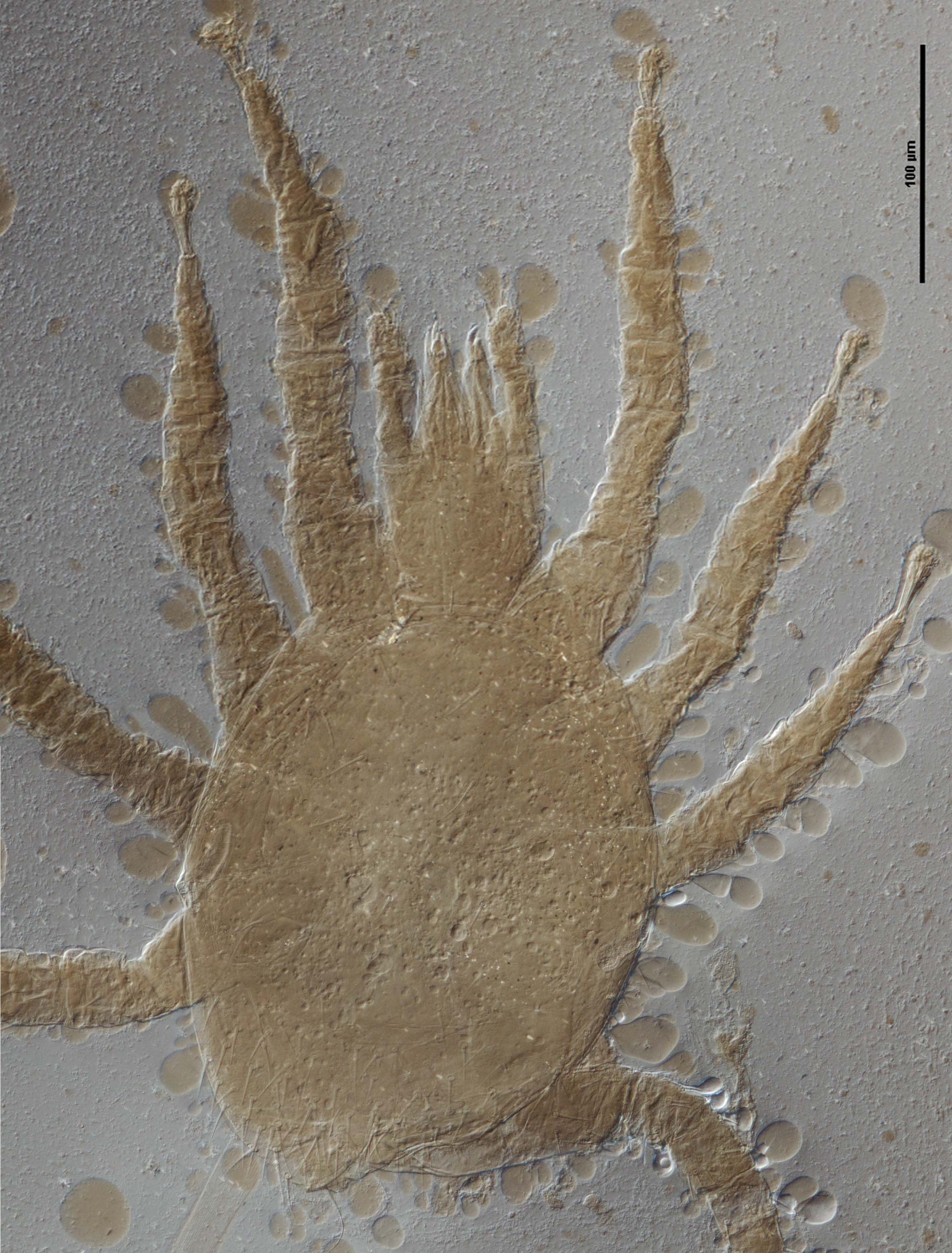
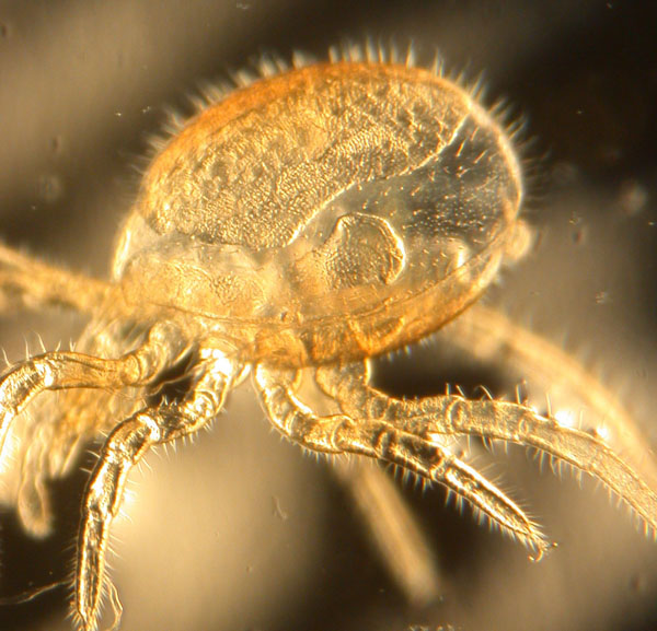
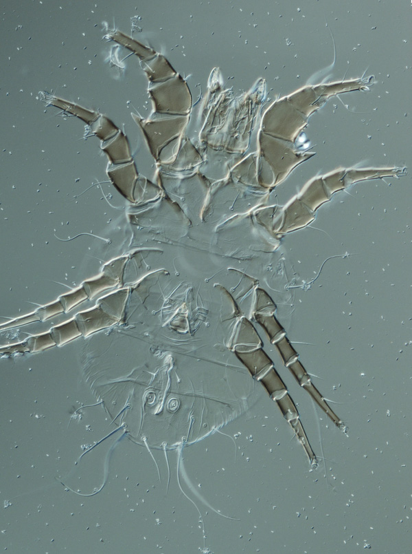
Male uropodid mite from Queensland, Australia

Berlese funnels

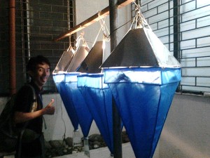
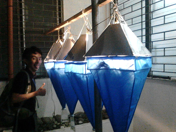
Berlese funnels at UPLB (photobombed)

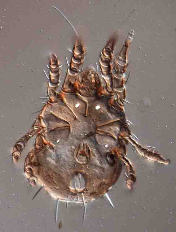
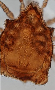
The group is the current focus of my research. They occur in all temperate and tropical areas of the world (there is a good diversity in Ohio), but some genera and families are restricted in distribution, usually to specific parts of the tropics. So off I went to collect in faraway places.
Mite collecting trips do differ a bit from classical big game collecting trips in Africa or India: 1) nothing is being shot, and 2) there is a noticeable lack of caravans of porters, elephants, etc. Most mite collecting involves either collecting directly from hosts (insects, vertebrates, etc), or from the habitat. For me habitat was the main target, given that most uropodines live in soil, litter, or rotting wood. Collecting mites is also not very glamorous and definitely lacks instant gratification: you go to a habitat, collect possible sources of mites (e.g. bags of soil & litter), and bring them back for processing. Processing usually means Berlese funnel extraction, using heat to drive the mites out of the substrate until they fall down the funnel and into preserving fluid (95% ethanol in most cases). Bottom line, you spend 30 minutes collecting, have to wait 2-3 days to see results, and many more weeks to figure out what exactly you got.  Patience is a virtue.  On the bright side, a single sample may yield hundreds of mites.

Mite collecting

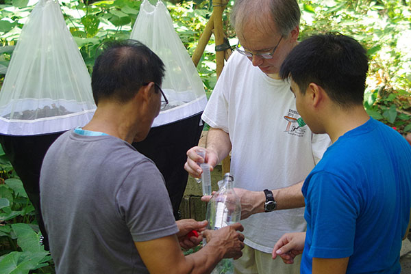
From left to right: Phin Garcia, author, Jeremy Naredo

The Philippines trip was standard in many ways, but exceptional in terms of scale. I usually come back from a trip with 2-8 samples, here we were running 20 funnels almost continually. It helped a lot to have good collaborators, and for this trip I was fortunate to be able to work with folks at the University of the Philippines Los Baños, the agricultural campus of the University of the Philippines. The help of Drs. Juan Carlos Gonzalez, UPLB Museum of Natural History director, and Jun Lit, director of the Arthropod collection, is greatly appreciated. I specifically worked with Jeremy (Jebboy) Naredo, a MSc student, and Rufino (Phin) Garcia, a staff member with an uncanny ability to find mites.

Our base of operations was the museum. The building is relatively small, but quite nice, and with small but extremely popular exhibit spaces. One day there were 8-10 buses of school kids parked in front.
The Museum is set in a remnant of tropical forest. To test the funnel assemblies we put up on the day I arrived, we grabbed some litter from around the building, and ended up with some of the richest samples of the entire trip. One more of the oddities of sampling for mites. The campus proved to provide some very good sampling opportunities, both around the museum and in the Hortorium, a much larger forest remnant along a creek running through campus.

Creek in Hortorium, UPLB campus

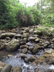
Creek running through UPLB campus

There are potential problems working in the Philippines. Collecting, even of litter samples, is strictly regulated, so we could only work in certain areas using permits issued to the museum. And as usual, not everything worked. A long anticipated trip to Sibuyan Island, the “Galapagos of the Philippines”, had to be cancelled because of rough weather. The only way to get to that island is by ferry, and with rough seas all ferry rides were cancelled. Disappointing, but there is a limit on how much risk to take to collect mites, and this was clearly too much.
The main trip was to the Laguna – Quezon Landgrant area, an area that has both decent forest and is the focus of additional reforestation efforts.

Laguna Quezon Landgrant area

Laguna Quezon Landgrant area

The trip there was interesting. Initially the standard fare, using a rented jeepney to get to the headquarters. After that it got more fun, when we climbed on a wagon towed by a tractor. I have strong suspicions that the folks at the station wanted to see how long we could last on a trip worthy of any amusement park ride. To say that the ride was “bumpy” is an understatement. We regularly went airborne, getting seriously worried about being flipped out of the wagon. Eventually we decided to walk the final part to save ourselves and the dissecting microscope I was bringing. On the way back we walked all the way with a horse carrying our supplies, much better. The camp we stayed at was basic, but by using a stream coming out of the mountains, there was running water and even nice (but cold) showers. And excellent yields of mites. Phin and Jeremy braved a colony of army ants to get some of the (temporary) nest material. The ants did not appreciate it, and later on tried very hard to leave the funnel through the top, instead of falling down in the alcohol. We had to tape up all access points to avoid having a colony in the museum.  The rangers in the area were great, although it was slightly disconcerting that they all carried shotguns (to protect against log poachers).

Mahogany plantation

Mahogany plantation

I never left the island of Luzon, but we did travel to the Northeastern corner of the island, a roughly 12 hr car ride. We went to visit Dr. Leonila Raros, the pre-eminent Philippine acarologist , who has retired there. The area is 95% rice paddy, but we sampled her bamboo plots, and a few other sites on the way. Samples included a mahogany (Swietenia macrophylla) plantation. This species of mahogany is native to Central America and increasingly rare in its native range. In contrast, it has become a pest plant in the Philippines, invading forest remnants and displacing native vegetation. Very little will grow under these trees in the Philippines

Cave at Pangasinan

Crew after sampling Pangasinan caves

Our final collecting trip was by far the dirtiest: caves at Pangasinan. The cave systems here are enormous, with a very active group of cavers constantly working on mapping the caves. I was interested because bat guano may house some very specific uropodines. 30 years ago I worked in the Philippines on a mammal survey, spending a lot of time in bat caves. All of those were dry, these were not. These cavers view of a “dry” cave was wading into water up to your knees, “wet” caves would require scuba gear (I only figured this out AFTER we visited). An interesting experience.

Overall we ended up with almost 400 vials of specimens, mostly uropodids. That may easily represent several thousand specimens. Does such level of collecting endanger species or destroy habitats? Almost certainly no. Each sample may represent about 2l (0.5gal) of soil and litter. Any road or housing construction will destroy far larger areas and mite numbers. Second, while most people think plants or vertebrates or butterflies when thinking about conservation, mites are parts of ecosystems, and to figure out what role they play it would be good to at least know what we have.

Thanks to Jeremy Naredo who provided most of the pictures.

About the Author: Dr. Hans Klompen is professor in the department of Evolution, Ecology and Organismal Biology and director of the Ohio State University Acarology Collection.

Tree holes and their mites

Many mites are very specific for particular habitats, whether it is the inside of the lip of a bat or a flower bud of a single plant species.  We have established this for many plant, insect or vertebrate associates. But knowing where exactly the mites are on a host is fairly easy. What about mites living is less discrete situations, like the litter layer? We are fairly sure that litter mites also have fairly specific microhabitats, but this is much more difficult to demonstrate.

image of tree hole sampled for mites   In the acarology lab we have been looking at one subgroup of litter habitats, tree holes. In this case we define tree hole as any cavity in the trunk of a tree that is not directly connected with the underlying soil. Tree holes in general may provide more stable microclimates, in terms of temperature, moisture, humidity and sun exposure, than standard litter habitats. All types of tree holes contain mites, and those mites tend to be specific to tree holes. Wet tree holes, containing water or just very wet litter, have been studied quite extensively because they are breeding grounds for certain species of mosquito, but we are particularly interested in dry tree holes. Initially we became interested in this microhabitat because we wanted to know more about distribution and habitat restrictions of Uropodella, a rare genus that had been found only in tree holes.  The genus Uropodella is most diverse in Chile, with only 1-2 species in North America. These mites appear to be phoretic on Tenebrionidae, and, fitting with that association, are found in very dry tree holes, containing nothing more than pulverized wood.

Image of grey squirrel

Gray squirrel

Mites in treeholes can also tell you something about other inhabitants of that treehole. Finding Aeroglyphidae in a S. Carolina tree hole indicated that there were probably bats roosting in that hollow tree, while the numerous Glycyphagidae in a tree hole in Columbus were consistent with the squirrel nest found in that same tree hole.

Glycyphagidae, ventral view of male

Glycyphagidae, ventral view of male

But this early research was largely anecdotal.

One of us, George Keeney, followed up in a big way by systematically sampling a large number of tree holes, some several times during different seasons.  The focus for this study was a quite diverse group, the Uropodina. We found that tree holes in Central Ohio not only have a quite diverse uropodid fauna, but that the species in tree holes tend to be tree hole specialists. A few species have been associated with a wide variety of tree species, while a number of other species have only been encountered only once or twice.  The two most commonly encountered tree hole species are Allodinychus nr. cribraria and Vinicoloraobovella cf.  americana. George affectionately calls the former species the “elf hat mite”, due to its fanciful resemblance to such!  That being said, we do not have enough information yet to determine whether these tree hole uropodines are specific for a given tree species, a given tree hole inhabitant (e.g. squirrels, birds, bats), particular exposure, tree hole size, tree hole litter moisture, season, etc., etc. To find out, we are now following up on the early survey, by recording more of these details, and especially by sampling many more tree holes.


Some tree species are more prone to developing dry tree holes in their trunks, usually at the site of branch removal or other injury.  Silver maples, magnolias, American beeches and American sycamore are some notable examples of such trees.  In Ohio, these species can be common in urban plantings and therefore, tree hole mite sampling can be quite productive in parks, campuses, street boulevards and other urban areas as opposed the more rural areas.  Such park trees are often mature and may have had large branches removed by landscaping and maintenance, providing the initial germ of many tree holes.  Sampling tree holes involves gathering the detritus from within the hole, though the hole should be thoroughly inspected before placing ones hands inside, as one may often be intruding upon the abode of a wary raccoon or testy gray squirrel!  So if you see anybody carefully trying to get “goo” out of a tree hole, it is not just fun, it might be research.

Tree hole in boxelder containing Philodana

Tree hole in boxelder containing Philodana

Philodana johnstoni, ventral view of female

Philodana johnstoni, ventral view of female

And there is always the option of finding something that is truly unexpected. One of the most spectacular for us was a hole in a broken branch of a box elder near campus at the Olentangy River Wetland Research park.  It contained a large population of Philodana johnstoni, a very odd species of Trigynaspid mite described from Ontario and New York, with no additional published records. It appears to be associated with the tenebrionid Neatus tenebrioides.

image of Don Johnston

Don Johnston

It is a very appropriate find, given the connection of this species with Ohio State University. Philodana johnstoni was named by John Kethley in honor of the previous director of the Acarology Laboratory, Don Johnston, and is a double honorific, as Philodana combines “philos” (=loving) and Dana, Don’s wife.


About the Authors: Dr. Hans Klompen is professor in the Department of Evolution, Ecology and Organismal Biology and Director of the Ohio State University Acarology Collection. George Keeney is Manager of the Acarology Collection and the OSU Insectary.

Pretty mite pictures


As a follow-up to my earlier blog post, I thought I should show some more examples or successes and problems in imaging mites.

Diplothyrus lecorrei (Holothyrida), nymph, single image using dissecting microscope

Diplothyrus lecorrei (Holothyrida), nymph, single image using dissecting microscope

The problem in the Diplothyrus image is depth of field.  The marginal areas of this mite are out of focus.

The LT-SEM image of Excelsotarsonemus shows what great depth of field can do.  Note the blobs of fungus stuck on this mite.  They are vectors of specific fungi.

Excelsotarsonemus sp. (Tarsonemidae), colorized LT-SEM

Excelsotarsonemus sp. (Tarsonemidae), colorized LT-SEM


Stacked imaging with Differential Interference Contrast illumination works very well with well sclerotized mites.  It generates nice images of cuticular patterns.


The technique can work well on poorly sclerotized mites, but in this case one has to worry about avoiding imaging structures from the other side.


Acarus siro, male, stacked image

Acarus siro, male, stacked image


The Trichocylliba images are a good example of what happens with stacked images to 3-dimensional structure.  These mites are as high as they are long, but the images do not show that.  Adding lateral images would solve it, but that proved very tricky with these mites.


Trichocylliba n.sp. (Uropodina), male, stacked image, 200x

Trichocylliba n.sp. (Uropodina), male, stacked image, 200x


Just for fun, a partial slice though the mouthparts of this Oudemansicheyla shows some of the power of Confocal microscopy (image courtesy of Sam Bolton and Gary Bauchan).


<i>Oudemansicheyla</i> (Cheyletidae), confocal microscopy

Oudemansicheyla (Cheyletidae), confocal microscopy


About the Author: Dr. Hans Klompen is professor in the Department of Evolution, Ecology and Organismal Biology and Director of the Ohio State University Acarology Collection.


Smile!! Imaging in the Acarology collection


Velvet mite, <i>Leptus</i> sp. (photo Rich Bradley)

Velvet mite, Leptus sp. (photo Rich Bradley)

Working with small organisms such as mites presents some interesting problems.  One of those is getting folks interested in just thinking about something they usually cannot see.  Of course a few mites are big enough to see and critters like ticks, velvet mites and water mites do enjoy some “popularity”.  But for most mites we have only fairly bad photographs of crushed specimens on slides (often good for seeing specific characters, but horrible for outreach).  Without nice images, we can still go for gross (I do admit that I occasionally enjoy going that route), with disgusting images of humans with scabies (Sarcoptes scabiei), or sad looking plants covered in spider mite silk, but it is not the same.  And it really does not do much for improving the image of mites.  Plus, most mites do not do any significant damage.  So how should we present the “real” mites?

Let’s start with the high-tech approach.

Scanning electron microscopy (SEM) was the first good option.  Because of the high depth of field, SEM gives very nice, 3-D images at very high magnification.  It works well for highly sclerotized mites (oribatid adults, uropodids) but it is less successful with small, soft-bodied species because most SEM techniques require critical point drying, which makes those soft bodied mites shrivel into ugly blobs.  Using live mites can alleviate that problem but (a) most SEM operators do not like it (it introduces humidity into the machine and may damage it) and (b) mites are often not cooperative.  As a beginning graduate student I had the option of putting some live sarcoptid mites in an SEM.  The specimens were on sticky tape and everything looked great as the SEM got close to vacuum.  However – there is always a “however” in these stories – just before we could get our first images the last of the great looking mites managed one more spurt of energy, and fell over, exposing nothing but its sticky stuff covered underside.  That took care of my hopes for great pictures.

Leg of <i>Osperalycus tenerphagus</i>, LT-SEM

Leg of Osperalycus tenerphagus, LT-SEM, colorized image

Low Temperature SEM (LT-SEM), uses liquid nitrogen to flash freeze the mites (no critical point drying), allowing views of live mites frozen in time.  This completely bypasses the shriveling problems of standard SEM with stunning results.  The folks at the USDA in Beltsville, MD, have generated some beautiful images of plant feeding mites.  Sam Bolton, graduate student in acarology, worked with them to get the images of his “dragon mite”.  Add some color (based on images of the mite taken before going into the SEM) and we end up with poster-worthy pictures.

Even LT-SEM has limitations, as we can only see surface structures.  We are overcoming that limitation by using confocal laser scanning microscopy.  With this technique you can also see internal structures, and with the right software, you can construct 3-D images that can be sliced any way you wish.  Not quite as detailed as Synchroton X-ray microtomography, but that technique requires a particle accelerator the size of a football field, and at this point only one (in Grenoble, France) seems to be set up to handle images of mites.  Confocal microscopy is great for research, but 3-D printing of the models also makes for excellent teaching tools (or gift store items).

A Confocal 3-D model of <i>Daidalotarsonemus</i> sp.  B 3-D print of the same

A Confocal 3-D model of Daidalotarsonemus sp. B 3-D print of the same

Of course all of those techniques require highly specialized and expensive equipment.  For day-to-day use we use an automated compound microscope and image stacking software to generate images that are both relatively detailed and retain some color.  These tools finally allow generation of well-focused detailed images of very small organisms and structures.  Unfortunately we do lose some of the 3-D effects in the process of image stacking, but on a good day, these images are publication quality.

For more hands-on displays, mites can be viewed on a 40 inch TV-screen using a video camera connected to a compound or dissecting microscope. This is wonderful for teaching (6-12 people can all watch what you are seeing in the microscope), research (sometimes the image on the TV is more detailed than the one you get through the eyepieces of the microscope itself), and of course outreach.  We use that set-up at the annual Museum Open House to show live mites in detail.

Mite on TV as used for teaching

Mite on TV

With enough technology, mites are coming out of the deep and dark, and into the light, where they of course belong.  We are slowly generating more high quality images and making them available through the collection database.  Check it out, the small can be beautiful.

About the Author: Dr. Hans Klompen is professor in the Department of Evolution, Ecology and Organismal Biology and Director of the Ohio State University Acarology Collection.


“(S)he got legs,” a look at mite legs


Mites are notorious for not following the “rules.”  For example, we generally teach that you can separate Insects from Arachnids by the number of legs, 3 pairs in insects, 4 pairs in arachnids. Simple, right?  Mites are arachnids, and most have 4 pairs of legs, but not all


And of course legs can get highly modified for various (known or unknown) purposes


About the Author: Dr. Hans Klompen is Professor in the Department of Evolution, Ecology and Organismal Biology & Director of the Ohio State University Acarology Collection.

The case(s) of the missing collection data


Sherlock Holmes silhouettePrevious blog posts have mentioned the value of data on collection dates, geographic localities, hosts (where relevant) and even things like the weather at the time of collection (collecting before or after a rain storm in the desert gives rather different results). For mites, the group I work on, we often want even more detail. Mites can be both very broad in their requirements and very picky. So information on microhabitat can be crucial. For example, in parasitic mites, we would like things like site on a host. If a mite was found in the quill of the secondary wing feathers of a house sparrow, I can probably give a pretty good guess on what it is. Not certain, there are always surprises, but I can make a good first guess. We know this by experience and by examining records in collections that have that level of precision.

In short, life is good as long as there are data, but what if you end up with specimens with little or no interpretable data? In the Acarology collection specimens are usually on microscope slides or in vials with alcohol.

image of good slides with poor data

Good slides with poor data

Many older slides or vials have labels giving only a partial identification plus a number or code, presumably referring to a notebook, letter, or other non-specified source. In principle fine, but without follow-up those notes get lost, misplaced, etc., and that means trouble. In one unfortunate case, roughly 3,000 chigger slides from South Korea, the data were deliberately omitted from slides and vials. These specimens were collected at the end of the Korean war, and it was probably not a good idea to specifically tell everybody where each military base was located. The chiggers were collected at that time and that place to help manage scrub typhus, a disease transmitted by chiggers. Good idea at that time, but it makes recovering the data at this time, sixty years after the armistice, quite tricky.

chigger photograph

A chigger in all its glory

Anyway, so what do you do when you have a bunch of microscope slides or vials with interesting specimens but no, or very little, data? That is where curation becomes a bit of detective work. There are some options.

Before and after microscope labels

Before and after microscope labels

1) If the specimens have at least a code/number, you often can find the same code of the same code format somewhere else in the collection. This is a brute-force method that requires databasing of large numbers of specimens. Luckily, the vast majority of the OSU Acarology Collection is now databased, with most label data captured. Find matching codes, hope that at least one slide has complete information, and if so, you are set for the other slides in the group. Even if the match is not perfect, a similar style of codes may help narrow down where poorly labeled specimens came from. For example, a certain style of code was used for specimens recovered from bats in Costa Rica. Starting with near complete data on 2-3 labels (out of ~400) and using an on-line mammal collection database (VertNet) we recovered all the relevant collection data and even know where the host specimens are located (Los Angeles County Museum in this case).

page from Lipovsky chigger notebook

Page from Lipovsky chigger notebook

2) Miscellaneous field note books, notes, etc. People have asked me why we keep file cabinets worth of old notebooks, notes, and letters from folks that left long ago. Barring proper log books, this may be where the data we want to recover comes from. One good notebook can solve problems with many specimens. Data for many of the Korean chiggers came from a notebook at the bottom of a drawer of largely useless paperwork. Of course it would be nice to have it all digitized, but we do not have the time, people or resources to do that at this time.

3) Sometimes it just takes some luck. For a long time I was going nowhere with a small set of slides with only identifications, and, on one slide, a name that seemed to refer to a locality in India, and something about a dung beetle. Then one day, out of the blue, I got an e-mail from somebody wondering whether I was interested in the remainder of a collection of mites he made from beetles in India. He had already sent me some specimens years ago (my mystery slides) and now I could add some real data. It does not happen very often, but it counts.

As a final note, some of you might think that the above does include quite a lot of interpretation. How accurate are these interpretations? This is a real problem. We try to be conservative in the “guesses” we make, but may fail on occasion. This is why we copy all original label data verbatim and make that available with each specimen. And we have had a few occasions where folks told us our interpretation was wrong. A big thank you to those folks!


About the Author: Dr. Hans Klompen is Professor in the Department of Evolution, Ecology and Organismal Biology & Director of the Ohio State University Acarology Collection. Silhouette of Sherlock Holmes: The Sherlock Holmes Museum. CC Attribution License. http://www.sherlock-holmes.co.uk. All other images courtesy of the author.