“Natural history collections are virtually inaccessible to everyone.”
This statement, in one form or another, can be found in many recent publications, online articles, opinion pieces, blog posts, and many comments on other social media outlets. To be absolutely honest, this irritates me greatly and there’s no better place to vent frustration than in a blog post!
Up until the late 1990’s the term ‘accessibility’ as applied to natural history collections, and insect collections in particular, had two basic components:
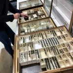
- Collection organization – with millions of specimens and many thousands of species, the material must be carefully organized, labeled, and catalogued so it is accessible when there is a request for information or for a loan.
- Services to scientists – scientists can access the specimens for the purpose of study, either through loans (upon request our staff selects, packages and ships specimens to scientists for study) or by visiting the collection.
Nowadays, we receive weekly requests that go more or less like this:
- Can I have the specimen data and images for each species of all your (name of insect group here)?
- Can you please take photos of the following (30 species) of (name of group here) for my (book, thesis, website, publication, database, etc.)?
Collections have quickly taken advantage of new computer and imaging technologies to provide new services to our user base, but with the advent of the Internet, browsers, most notably Google, and now mobile technology, collections are facing new, and I argue sometimes very unrealistic, expectations of services by our existing users, new users, and even funding agencies.
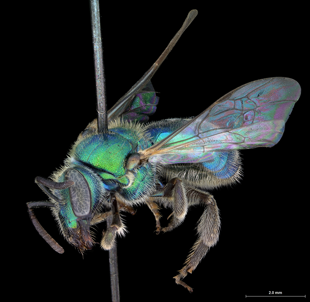
Augochlorella pomoniella OSUC 128046
There is a strong and fast-growing demand for high resolution images of specimens and of specimen label data that can be easily plugged into studies of global climate change, evolutionary biology, conservation, etc. Please don’t get me wrong! This is awesome! We have been saying for many years that collections are an enormous resource of precious information and we stand by it! However, this relatively recent demand did not come with funds to support much needed basic collection curation or for hiring and training of permanent curatorial staff.
‘Accessibility‘ today goes way beyond old-fashioned physical access to specimens or even online catalogs. It includes the expectation (and demand really!) of having all the specimen level data captured and remotely accessible now. Collections continue to incorporate new technologies into our curatorial protocols to provide the best service we can to our users. But what is reasonable? What’s possible, particularly with the reality on the ground?
Collection curation (= maintenance and improvement) takes 1) people, 2) time, and 3) money. Maybe one day technology will eliminate the need for humans handling collection specimens, but that does not look very likely in the near future. In the meantime most collections are chronically underfunded. In our case, we do not have a centrally supplied operating budget.
In university settings, most of our work force are undergraduate students and, as a consequence, highly temporary in nature. No matter how smart and dedicated our undergraduate students are, they usually have no prior curatorial experience and must be trained from scratch. The constant training and management of part-time workers is very time-consuming and stressful for the few permanent staff.
Extra-mural funding options for collections are limited to donations, and, on occasion, grants for special projects. Our day-to-day operations (specimen preparation, loan-related activities, visitor support and infrastructure, and much more) are basically self-funded and therefore done if and when we have money, personnel and time to do it.
We at the Triplehorn Insect Collection continue to be committed to making collection information available online. We were one of the first insect collections in the world to make specimen data remotely available: our website has been on-line since 1994, and dynamic access to specimen data since 1997. Our state-of-the-art web interface serves all but one of the collections here at the Museum of Biological Diversity as well as various partner institutions across the country and abroad.


Our level of commitment, however, is running up against the limits of our resources. We’re trying to think of new and productive ways to generate financial support, and as much as we dislike it, everything is on the table. Some museums charge “bench fees” to visitors (we don’t yet); perhaps we should consider a virtual bench fee. Can the data and images that we create be monetized in other ways as well, particularly for commercial purposes? So far all our data have been made freely available online, and we like it that way. But how long will we be able to afford it? This is all very complicated and made more difficult by the fact that we work within the context of a taxpayer-supported public university.
Speaking of taxpayers, the general public can help collections by supporting our efforts. Volunteers, donations (our Friends of the Triplehorn Insect Collection fund is 100% used for the care of the collection), and just word-of-mouth are all critical to the cause.
More information about the Triplehorn Insect Collection is available on our (soon to be revamped) website. The collection Facebook page brings recent updates & fun stories. Also, check out our collection blog, Pinning Block, for a view of the collection, our people and the work we do.
About the Authors: Dr. Luciana Musetti is Curator of the Triplehorn Insect Collection at Ohio State University. Dr. Norman F. Johnson is Director of the Triplehorn Insect Collection and Moser Chair in Arthropod Systematics and Biological Diversity in the Department of Evolution, Ecology, & Organismal Biology and Department of Entomology at The Ohio State University.
Follow us, @osuc_curator & @baeus2 on Twitter for our more personal views & commentary.