Further to my recent blog on confocal microscopy, here are some images of 3D models that I generated using a confocal laser scanning microscope.
-

- Many mites, including this alycid (Pachygnathus), have pincer-like chelicerae for breaking into food items.
-

- Here is a rather peculiar-looking mite (Daidalotarsonemus) that was collected from the surface of leaves from a Brazilian rainforest.
-
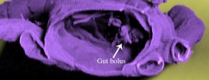
- Here is the inside of a mite (Stigmalychus), viewed from behind. There is a readily visible gut bolus inside the mite in addition to other pieces of food.
-

- Some mites, such as this alycid (Pachygnathus), have eyes. This image shows a lateral view of the front of the mite.
-

- And just in case you’re getting bored of mites, here’s a symphylan (myriapod).
-

- Occasionally a confocal microscope can lead you to produce artistic or aesthetically pleasing images. This is a ventral view of the mouthparts of a plant-feeding mite – Brevipalpus. The surface of this mite reminds me of folding fabric.
Acknowledgements: The 3D models were generated at the Beltsville Agricultural Research Center, US Department of Agriculture. The Daidalotarsonemus specimen was provided by José Marcos Rezende, São Paulo State University, Brazil.
About the Author: Samuel Bolton is a PhD student at the Acarology Laboratory at the Museum of Biological Diversity. He regularly uses confocal microscopy for his research on the mouthparts of mites.
Intriguing images and nicely depicted Sam, well done!
Thanks Marc