Explaining Science – vermiform mites

You have heard of mites – minute arachnids that have four pairs of legs when adult, are related to the ticks and live in the soil, though some are parasitic on plants or animals. But what are vermiform mites? Maybe you have heard of vermi-compost, a composting technique that uses worms (like your earthworm in the garden) to decompose organic matter. So vermiform mites are mites with a body shape like a worm:

worm-shaped nematalycid Osperalycus

Why are they shaped like a worm, you may ask – To find out more I interviewed Samuel Bolton, former PhD student in the acarology collection at our museum, now Curator of Mites at the Florida State Collection of Arthropods. Sam’s main research interest is in mites that live on plants and in the soil, especially Endeostigmata, a very ancient group of mites that dates back around 400 million years, before there were any trees or forests. Sam’s PhD research with Dr. Hans Klompen here at OSU, was focused on a small family (only five described species) of worm-like mites, called Nematalycidae.

side note: You may have heard of Sam’s research in 2014 when he discovered a new species of mite, not in a far-away country, but across the road from his work place in the museum.

When Sam started his research it was not clear where these worm-like mites in the family Nematalycidae belong in the tree of life. To find out Sam studied several morphological characters of Nematalycidae and other mites. He focused in particular on the mouth-parts of this group. As he learned more about the mouth-parts of this family, he found evidence that they are closely related to another lineage of worm-like mites, the gall mites (Eriophyoidea). Eriophyoidea have a sheath that wraps up a large bundle of stylets. They use these stylets to pierce plant cells, inject saliva into them and suck cell sap.
Although Nematalycidae don’t have stylets, one genus has a very rudimentary type of sheath that extends around part of the pincer-like structures that have been modified into stylets in Eriophyoidea.

So what did Sam and his co-authors discover?

“.. Not only are gall mites the closest related group to Nematalycidae, but the results of our phylogenetic analysis places them within Nematalycidae. This suggests that gall mites are an unusual group of nematalycids that have adapted to feeding and living on plants. Gall mites use their worm-like body in a completely different way from Nematalycidae, which live in deep soil. But both lineages appear to use their worm-like bodies to move around in confined spaces: gall mites can live in the confined spaces in galls, under the epidermis (skin), and in between densely packed trichomes on the surface of leaves;  Nematalycidae live in the tight spaces between the densely packed mineral particles deep in the soil.”

This research potentially increases the size of Sam’s family of expertise, Nematalycidae, from 5 species to 5,000 species. We have yet to confirm this discovery, but it is highly likely that gall mites are closely related to Nematalycidae, even if they are not descended from Nematalycidae. This is interesting because it shows that the worm-like body form evolved less frequently than we thought. This discovery also provides an interesting clue about how gall mites may have originated to become parasites. They may have started out in deep soil as highly elongated mites. When they began feeding on plants, they may have used their worm-shaped bodies to live underneath the epidermis of plants. As they diversified, many of them became shorter and more compact in body shape.

I wish I could tell you now to go out and look for these oddly shaped mites yourself, but you really need a microscope. Eriophyoid mites are minute, averaging 100 to 500 μm in length. For your reference, an average human hair has a diameter of 100 microns.

eriophyoid Aceria anthocoptes

Reference:

Bolton, S. J., Chetverikov, P. E., & Klompen, H. (2017). Morphological support for a clade comprising two vermiform mite lineages: Eriophyoidea (Acariformes) and Nematalycidae (Acariformes). Systematic and Applied Acarology, 22(8), 1096-1131.


About the Authors: Angelika Nelson, curator of the Borror Laboratory of Bioacoustics, interviewed Samuel Bolton, former PhD graduate student in the OSU Acarology lab, now Curator of Mites at the Florida State Collection of Arthropods, in the Florida Department of Agriculture and Consumer Services’ Division of Plant Industry.


Squirreling in the Pacific Northwest

You may have heard that researchers discovered a new species of flying squirrel. These squirrels had lived in plain sight for decades but only recently did Brian Arbogast and colleagues investigate the DNA of some of these animals. Their findings were revealing: The Pacific squirrels cluster separately from the northern and southern flying squirrel. The researchers analyzed mitochondrial DNA as well as microsatellite data to reveal this new evolutionary relationship.

Note: Mitochondrial DNA and microsatellites are parts of a species’ genome that are regularly used to construct evolutionary trees. In addition to the DNA in every cell’s nucleus in our body, mitochondria, the energy powerhouses in our cells, have their own genome. This mitochondrial genome is relatively small, is inherited from the mother only and has relatively high mutation rates. It is like a small clonal lineage within an organism which makes it ideal for evolutionary studies.   Microsatellites are short sequence repeats in the nuclear genome that do not produce proteins. Thus they are free to mutate at a higher rate than coding sequences – mutations will not mess up protein production- and they frequently vary in length and thus reveal relationships among organisms. 

A few weeks ago, before this study was published, 2 species of flying squirrels were considered to exist in North America, the northern and the southern flying squirrel. Here in Ohio the northern flying squirrels is resident – it is nocturnal though, that’s why you probably have not seen one yet.

Map showing distribution of now 3 species of flying squirrels

Map showing distribution of now 3 species of flying squirrels

DNA analysis showed that the coastal squirrels in Washington and Oregon are distinct from their northerly relatives and that they actually only co-occur with them at 3 sites in the Pacific Northwest. Northern and the newly described Humboldt’s flying squirrel do not interbreed at these sites. By the way, the researchers named the new species Glaucomys oregonensis because the specimen that was used to describe the species was collected in Oregon.

You may recall from a previous post, that Dr. Andreas Chavez in our department of EEOB studies relationships among squirrels in a different genus, Tamiasciurus, the red squirrel T. hudsonicus and the Douglas squirrel T. douglasii. These two species share habitat in the Pacific Northwest and they do hybridize.

Dr. Chavez was not available for an interview for his thoughts on the new species description of flying squirrels, because he is currently pursuing his own fieldwork in the Pacific Northwest. He and his field assistant Stephanie Malinich are collecting data to better understand the hybrid zone dynamics between the Douglas and red squirrel.

We will give you an update on Dr. Chavez’ research once he returns.

About the Author: Angelika Nelson is the curator of the Borror Laboratory of Bioacoustics and writing this post for Stephanie Malinich, collection manager of the tetrapods collection. Stephanie is currently doing fieldwork on the red and the Douglas squirrel in the Pacific Northwest.

Why describing new species is exciting and important!

For many researchers describing a new species seems like a tedious task. The differences between species might not be obvious, and the language confusing and foreign. This fact became apparent to me when I first presented my work to the Ant Lab at the Museum of Biological Diversity (MBD). As I described subtle differences in morphology, a little spine here and the shape of a hair there, I could tell that I had lost my audience by the dulled looks on my lab mates faces. How could they not see the differences in these two species?

comparison of Trachymyrmex new species and T. zeteki

Fig. 1 – Trachymyrmex new species on the left and T. zeteki on the right

“Some key differentiating characters: The integument is granulose, spatulate bi-colored setae occur between the frontal carina, the scape extends past the occipital corners. This is compared to a weakly irrorate integument, simple bi-colored setae between the frontal carina, and the scape reaching the occipital corners.”

Fig. 2 – In case you are not familiar with the some terms used in describing ant species


Totally clear, right?

While the differences in characters that separate Trachymyrmex new species and T. zeteki, are exciting for me, it seems to bore people to death. After my presentation, I received very helpful constructive criticism from my lab group. They thought it was interesting but a lot of my presentation went over their heads. My advisor, Dr. Rachelle Adams (Assistant Professor in the Department of Evolution, Ecology and Organismal Biology), encouraged me to find a way to turn the jargon into something people can digest and appreciate. I am still working on that, and it is a challenge many researchers face.

Species descriptions are important and a necessary part of daily life

Hopefully your parents told you when you were younger, never eat mushrooms you find in the woods. Taxonomy helps us understand what kind of mushroom you found, if it is edible, or if it might seriously hurt you if you eat it. Mushrooms are a great example of why taxonomy is important. Scientists need to describe and name species so that others can learn which characteristics define a species. Then chemists can tell us which are toxic. This information communicated to the public can potentially save lives! Taxonomists donate representations of species in museums so that they can be compared by other scientists in the future. Aside from publishing their species description, they submit the specimen used to describe the new species, a type specimen. Anyone who works with any type of animal or plant should be submitting voucher specimens, physical specimens that serve as a basis of study, as representatives of their work.

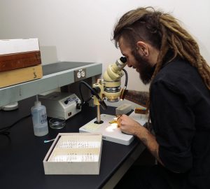
Cody working at microscope

Fig. 2 – Photo courtesy of Plain Janell Photography

My Taxonomic Conundrum

While working on my species description, I reviewed all the literature that included T. zeteki. The 30 papers covered a number of areas such as fungus-growing ant genomes, mating systems, alarm pheromones, larvae development, and gut bacteria. Sadly, almost half of the papers do not mention depositing voucher specimens! Two articles deposited their DNA sequences as vouchers to a database for molecular data. Any research that uses DNA sequences has to submit DNA vouchers to that database; without it your work cannot get published. However, they do not have any physical vouchers linked to their sequences! This lack of physical vouchers was quite a surprise to me. The time I spent as an intern at the MBD Triplehorn Insect Collection, my advisors and other mentors strongly advocated the deposition of vouchers. Without being able to link your DNA sequence to a correctly identified organism, that DNA voucher loses its value. You cannot quickly identify an organism from DNA. Using morphology is the easiest way to do so! It seems many researchers don’t recognize the importance of vouchering and most non-taxonomic journals do not demand it. Research published without vouchers lacks reproducibility, an essential component of the scientific method.

In my research project, I am cleaning up the mess left behind from nearly twenty-years’ worth of poor vouchering and misidentification. I’m not only describing a new species and key characters that differentiate two cryptic species, I am listing all of the papers that have been published in the past twenty years using the names Trachymyrmex zeteki and Trachymyrmex cf. zeteki. By linking the new species description to these articles scientists can move forward knowing the proper identification of these hard-to-identify fungus-growing ants.

The deposition of vouchers should be required for all publications, and is crucial for, past, present, and future research in biology. In my undergraduate research, I discovered there is a disconnect between research museums like the MBD and many scientists. While I am still struggling to turn the technical jargon into information that can be swallowed by non-experts, there are discussions to be had about the importance of taxonomy as a cornerstone in biology.

If you want to learn more about fungus-growing ants and the importance of university research collections, come see us at the MBD Open House April 22, 10am – 4pm.

CodyCardenas, undergraduate student ant lab, EEOBAbout the Author: Cody R. Cardenas is a Senior Undergraduate student in Entomology  working in the Adams Ant Lab.

*** We would like to hear from you – Please leave a comment ***