A Snapshot of Ohio Lichen Diversity 125 Years Ago

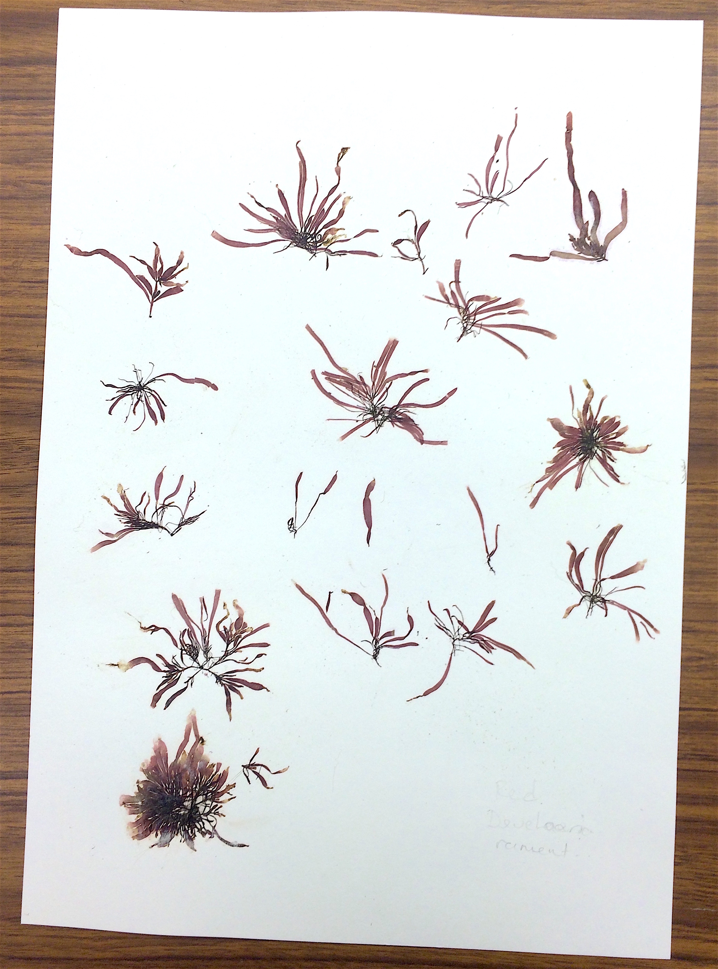
The Kellerman Displays for the 1903 Chicago Exposition

Most of the specimens at the Ohio State University Herbarium (OS) are tucked neatly into cabinets, not on display. But adorning one long wall are what at first glance look like pictures. Artfully arranged, with wood frames and a glass front, a close look reveals they are not paintings but are in fact real, once-living, plants and fungi.

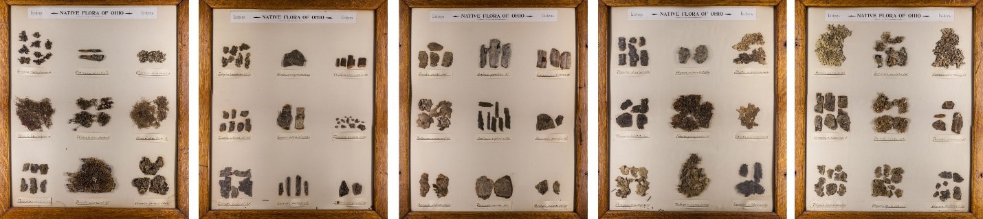
Framed specimens at The Ohio State University Herbarium

The displays are quite pretty and they’re obviously rather old, but I never stopped to consider just how old they are, or how they came to be. A modern interpretive sign explains that they, along with four larger, more intricate panels of Ohio trees, were assembled for display at the World’s Columbian Exposition, a big world’s fair held in Chicago for six months in mid-1893.

write-up by Ronald L Stuckey about Kellerman's Columbian exposition mounts

Write-up by Ronald L Stuckey about Kellerman’s Columbian exposition mounts

At the top of each 18 x 22-inch panel is a printed heading “Flora of Ohio,” and beneath that, in ornate old-style penmanship, are the words “Prepared by Professor and Mrs. W. A. Kellerman.” William A. Kellerman was remarkably energetic and wide-ranging in his botanical interests. Making these panels was an appropriate hobby for a person whose life revolved around plants and fungi. An Ohio native born in 1840, he attended Cornell University for undergraduate studies and later received his Ph.D. from the University of Zurich, Switzerland. He taught at schools in several nearby US states before returning home to become OSU’s first botany professor and Chairman of the Department of Botany when it was formed in 1891. That same year, he established the Herbarium in a building aptly named “Botany Hall” that unfortunately no longer exists on OSU’s oval. Since then the Herbarium has moved twice, first to the also aptly named “Botany and Zoology” building (now Jennings Hall) and then to its present location as part of the Museum of Biological Diversity on West Campus (1315 Kinnear Rd.). While his principal research interest was rust fungus diseases of crops, Kellerman’s numerous works on the flora of the regions where he lived reveal an extraordinary breadth of knowledge. He wrote a guide intended principally for use by teachers entitled “Spring Flora of Ohio” (1895) and co-authored, beginning in 1894 and subsequently updated several times, “A catalogue of Ohio Plants.” Sadly, while Kellerman was on a research trip to study fungi in Guatemala, he contracted a fever (most likely malaria) from which he died in 1907.

Photo of WA Kellerman in the Journal of Mycology

Photo of W.A. Kellerman in the Journal of Mycology

The panels are an interesting snapshot of the flora of Ohio. While aesthetics and enthusiasm for particular plants may have played a major role in their selection by the Kellermans, the panels were indeed portrayed to fairgoers as indigenous representatives of our flora. As there have been substantial changes in the composition of our vegetation, especially for such pollution and disturbance-sensitive organisms as lichens, they arouse curiosity about the past versus present status of these organisms.

Lichens are dual organisms consisting of fungus plus alga. The algae are single-celled photosynthetic organisms. The fungus, which constitutes most of the body of a lichen, provides a home for the algae, usually in a layer just beneath the surface. Most lichens fall into one of three growth-form categories: (1) usually small “crustose” lichens that are tightly attached to the substrate and so don’t have a discernable lower surface; (2) small to medium-sized “foliose” lichens that are flattened and can usually be separated from the substrate, and (3) “fruticose” lichens that have a bushy shape, either standing upright from the surface they are growing on, or dangling off a tree branch or trunk. Most of the lichens in the panels are foliose species.

Illustration of three growth-form categories of lichens

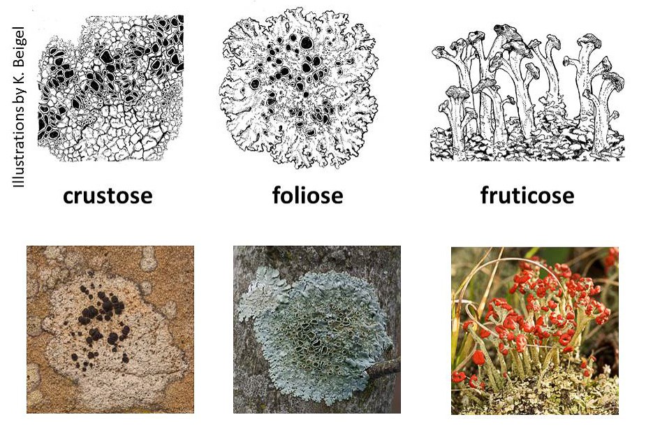
Three growth-form categories of lichens

There doesn’t seem to be a strict organization scheme for the lichen panels; they’re not in alphabetical or taxonomic order, except that one panel consists mostly of crustose species, while the few fruticose ones represented are grouped together, sharing space with some foliose ones. I suspect that the paucity of fruticose types is attributable to the display method only being suitable for flat or readily flattened specimens.

Each panel includes 9 specimens, with handwritten labels. The classification of lichens has undergone substantial change in the past century and a quarter, hence many of the names written by the Kellermans are not in use today. Fortunately, an on-line database called “Consortium of North American Lichen Herbaria” lists specimen records for lichens residing in collections spanning the continent, and the site lists all the names by which a species has been known in the past.

The present distribution of lichens in Ohio is well described in The Macrolichens of Ohio by Ray E. Showman and Don G. Flenniken, published in 2004 by the Ohio Biological Survey, and distribution maps presented on the web site of the Ohio Moss and Lichen Association. The status of the lichens more broadly is set forth in a monumental book, Lichens of North America by Irwin M. Brodo, Sylvia D. Sharnoff and Stephen Sharnoff, published in 2001 by Yale University Press, along with an updated companion volume by Brodo published in 2016 by the Canadian Museum of Nature, Keys to Lichens of North America: Revised and Expanded.

One panel caught my eye. This is a group of mostly rather large foliose lichens, including several “lungworts,” members of the Lobaria –robust broad-lobed species found on bark.

Display of a group of mostly rather large foliose lichens

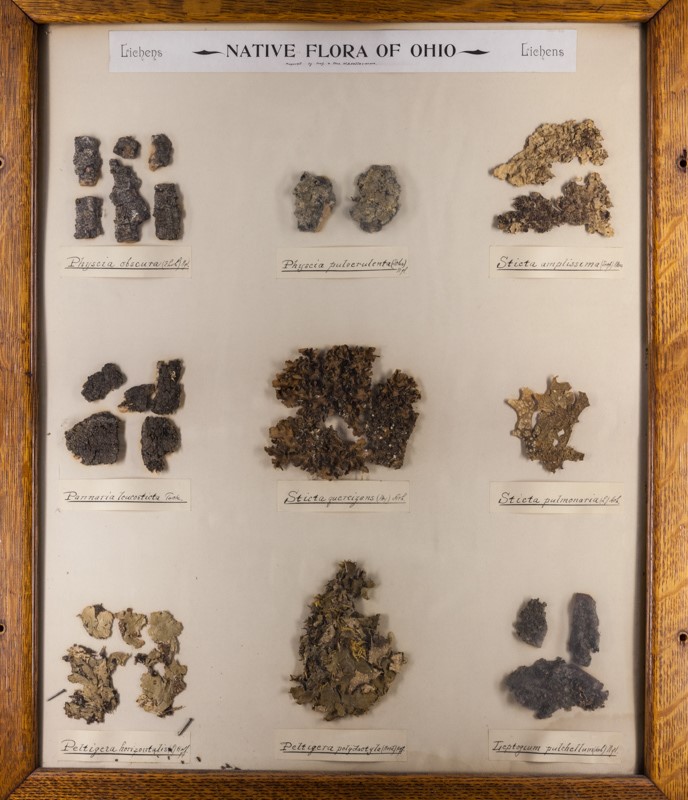
A group of mostly rather large foliose lichens

Among the most easily recognized of all lichens, lung lichen, Lobaria pulmonaria, was once widely distributed across Ohio, but no more. All but one of the 14 county records for lungwort are pre-1945, with the other one record being sometime between 1945 and 1965. Extensive searching has failed to find lung lichen today.

Why is it lung lichen gone from Ohio? It’s probably due to a multiplicity of factors that prevailed during the late 19th, and early 20th centuries: air pollution and disturbance of old-growth forests. Now that conditions are better for it to grow, perhaps a lack of propagules is keeping it from reestablishing itself. While eventually a warbler or vireo might fly in from some north woods with a little piece of lungwort on its foot, this might be a good candidate for a deliberate reintroduction.

Photo of lungwort growing on a tree in Maine

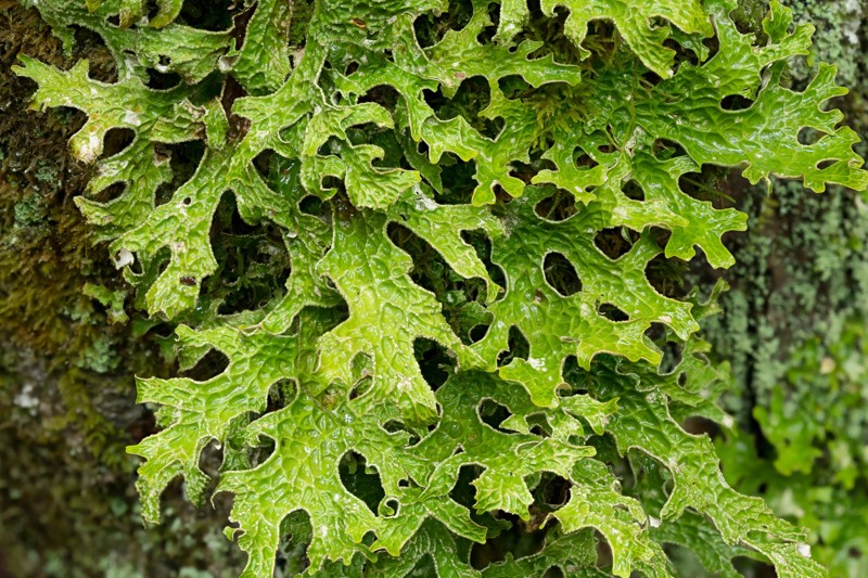
Lungwort growing on a tree in Maine

This is what lungwort looks like, growing on a tree in Maine. It’s a beautiful lichen and that just might still be growing in in a bottomland forest somewhere in Ohio, or it might soon return. Keep an eye out for it the next time you go hiking!

About the Author: Bob Klips is Associate Professor Emeritus in the department of EEOBiology at The Ohio State University. He currently assists with moss and lichen databasing in the OSU herbarium. His research focuses on bryophyte ecology.

Meet the Seaweeds

In this post, I would like for you to meet some of the algae that are to be placed in the OSU herbarium that I collected and processed into specimens from shorelines close to Eagle Hill Field Station in Maine, July 2016 (see below).

This slideshow requires JavaScript.

During part of the class, students were introduced to the ethnobotany of algae, i.e., human uses for seaweeds. Brown algae usually contain a polysaccharide, algin, in their cell walls. Algin is used in industry, food and pharmaceuticals to thicken and stabilize products. Examples of products that contain algin include ice cream, yogurt, soups and sauces, medicine tablets and antacids, toothpaste, lotion, lipstick and paint.

Below is some ethnobotanical information that I learned about the species of algae in the slideshow above.

Nori is an edible seaweed that dries to paper-thin sheets. It is often used as a wrap in sushi. In Maine, nori is currently harvested from wild-grown algae beds, however, nori farming soon will be a reality off the coast of Maine.

Irish moss is a primary source of carrageenan that is used as a thickening agent in toothpaste, foods and drinks, such as ice cream, pudding, and beer. The arrow in the photo points to round cystocarps that produce reproductive spores that will be released into the water when mature.

The winged kelp specimen was attached to a rock on the sea floor about four feet below water level at low tide. Winged kelp is edible and it is used in salad, soups and miso. It has a nutty flavor and is high in vitamins.

Dulse is added to salads, sandwiches and soups, and it is also eaten as chips. It is high in vitamins, minerals and amino acids.

Bladder wrack (don’t you just love the name) grows in extensive mats covering the tops of rocks above the low tide line. It is added to soups, applied medicinally to prevent skin irritations, used as a sunscreen, or utilized as a fertilizer. Like many edible seaweeds, it is high in minerals and vitamins. The paired bladders found at the ends of the branches (arrow on the bladder wrack photo) contain a gel with reproductive spores inside. The brown coloring around the bladders (see above) is algin that leached out of the bladders into the surrounding paper.


To make the herbarium specimens shown above, the seaweeds were processed and pressed in the lab after collecting. Preparing seaweeds for pressing is more elaborate than for land plants and often can be an artistic process requiring time and patience.

Since seaweeds are thin and fragile, they often break apart and/or stick together when removed from the water. Therefore, seaweeds are “floated” onto herbarium paper before pressing so that they can be arranged as close to their natural state as possible.

“Floating” is accomplished by placing a herbarium sheet under the water (water from the sea) with the seaweed floating on the water surface. The paper is carefully lifted up while the water slowly drains off the paper leaving behind the algae in a relatively natural position on the paper. However, slowly draining the water may not sufficiently separate smaller branches of the seaweed. In this case, the branches need to be painstakingly teased apart with a blunt probe while a thin film of water is still present on top of the paper.

Below, meet two additional species of seaweeds that I collected that are finely branched, and that required significant time and an artistic flare to arrange and separate tiny branches.

A finely branched red alga.

A finely branched red alga.

As of yet, the red alga on the left is unidentified, but I know it is polysiphoneous which means it has a central core of cells surrounded by a cortex. The branches appear jointed because the cells of the core and cortex are the same length, beginning and ending at the same place. This species also has tiny spines at each joint.

A brown alga that is finely branched in a feather-like pattern, possibly in the genus, Desmarestia.

A brown alga, possibly in the genus, Desmarestia.


Each side branch of the brown alga on the right was carefully separated from the main branch using a blunt probe to gently nudge the branches into position as the plants were “floated” onto herbarium paper before drying.


After pressing and drying, some seaweeds need to be glued onto the paper and some, especially brown algae that contain algin, often adhere to the herbarium paper without glue. Some algae, such as the finely branched red algae, make exquisitely beautiful specimens.

Meet a couple more unique and beautiful brown and red algae species collected in Maine near Eagle Hill Field Station (below). Note the brown shadow around the sausage weed specimen. The shadow is formed from algin that diffused from the alga into the surrounding paper.

This slideshow requires JavaScript.

In closing, I will leave you with a fun fact about sausage weed. The name sausage weed comes from the branches that have sausage-like constrictions. Trivia time, here we come!


About the Author: Dr. Cynthia Dassler is curator of Cryptogams (small plants that produce spores) at The Ohio State Herbarium (OS) in the Department of Evolution, Ecology and Organismal Biology.

Photos by Cynthia Dassler.


See the Seaweeds

As a scientist and curator, learning is a constant endeavor. Mix this with the continuous increase in knowledge about organismal relationships and classification, and training becomes critical to maintaining knowledge necessary to effectively curate collections in the OSU herbarium. Thus, I traveled, sixteen hours by car, to Eagle Hill Field Station in Steuben, Maine at the end of July 2016 to attend two, week-long courses, one of which was “Introduction to Maine Seaweeds: Identification, Ecology, and Ethnobotany.”

My reasons to take a course about seaweeds were two-fold: the OSU herbarium has a very small collection of seaweeds, or macro-algae, and I wanted to add to it. Secondly, I had very little experience collecting seaweeds, and thus sought to learn proper collection and curating techniques.

During the class, we spent about three hours each day collecting seaweeds at low tide. We collected along different types of shorelines near the Field Station.

Shorelines with large rocks and ledges are common near the Field Station (see below). The dark rocks closer to the water are covered with brown rock weeds. Large rock ledges occur at the water’s edge. At low tide near the rock ledges, the water is about chest high. Many different species of seaweeds, especially red algae, occur beneath the water surface attached to the sides of the large rocks. Large brown kelps, such as sugar kelp and winged kelp, are underwater and attached to rocks on the sandy bottom.

A typical rocky shoreline at low tide along the Maine coast near Eagle Hill Field Station.

A typical rocky shoreline at low tide along the Maine coast near Eagle Hill Field Station.

Rock weeds often cover the rocks from the high tide line to almost the water’s edge. The rock weeds are usually of two species: knotted wrack (Ascophyllum nodosum) and bladder wrack (Fucus vesiculosus). Both species are shown in the slide show below. Bladder wrack can be identified by paired air bladders (green bumps on the plant on the left side of the close-up photo of rock weeds). Knotted wrack possesses singularly arranged air bladders along the branches (inflated areas on the plant on the right side of the close-up photo of rock weeds). The creamy white bumps on the rock surfaces are barnacles that cover nearly every rock that receives water at high tide. Pools among the rocks are often havens for red algae, small shrimp, crabs and sand dollars.

This slideshow requires JavaScript.

What an incredible experience! Where else could I spend an afternoon clambering over barnacle-covered rocks, poking my hands into pools and crevices looking for red, brown and green seaweed treasures.

A different type of shoreline that we visited is characterized at low tide by open, exposed sandy or gravelly bottoms that often have scattered rocks (see slideshow below). The water is often cloudy, and seaweeds that are attached to rocks beneath the water are difficult to see. The students were often seen strung along the shore, hoping to find a unique specimen in the cloudy water.

After collecting, we began the elaborate task of processing and pressing the seaweeds to create archival specimens by “floating” them onto herbarium paper before drying them in a plant press. On a photo in the slideshow below, one of the students is holding a jug with seawater. We used seawater in the lab to “float” the algae onto the herbarium paper. Tap water is not ever used, as it kills the algae and makes the specimens mushy.

This slideshow requires JavaScript.

This is where I leave you until next time. Next time, look forward to meeting some of the seaweeds that I collected, but here is a teaser:

collection sheet of red alga, Devaleraea ramentacea

A collection of the red alga, Devaleraea ramentacea.

Above, each individual alga was “floated” and arranged on a herbarium sheet before drying in a plant press. The specimen was found underwater attached to rocky ledges that were partly exposed at low tide. I collected this alga by reaching underwater and feeling along a rock, choosing it because it felt different than the other algae around it. This species has a limited native distribution, found only along the northern Atlantic coast from Cape Cod to Labrador and Newfoundland.

About the Author:  Dr. Cynthia Dassler is curator of Cryptogams (small plants that produce spores) at The Ohio State Herbarium (OS) in the Department of Evolution, Ecology and Organismal Biology.

All photos were taken by Nick Blouin.