Smile!! Imaging in the Acarology collection


Velvet mite, <i>Leptus</i> sp. (photo Rich Bradley)

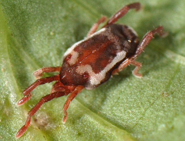
Velvet mite, Leptus sp. (photo Rich Bradley)

Working with small organisms such as mites presents some interesting problems.  One of those is getting folks interested in just thinking about something they usually cannot see.  Of course a few mites are big enough to see and critters like ticks, velvet mites and water mites do enjoy some “popularity”.  But for most mites we have only fairly bad photographs of crushed specimens on slides (often good for seeing specific characters, but horrible for outreach).  Without nice images, we can still go for gross (I do admit that I occasionally enjoy going that route), with disgusting images of humans with scabies (Sarcoptes scabiei), or sad looking plants covered in spider mite silk, but it is not the same.  And it really does not do much for improving the image of mites.  Plus, most mites do not do any significant damage.  So how should we present the “real” mites?

Let’s start with the high-tech approach.

Scanning electron microscopy (SEM) was the first good option.  Because of the high depth of field, SEM gives very nice, 3-D images at very high magnification.  It works well for highly sclerotized mites (oribatid adults, uropodids) but it is less successful with small, soft-bodied species because most SEM techniques require critical point drying, which makes those soft bodied mites shrivel into ugly blobs.  Using live mites can alleviate that problem but (a) most SEM operators do not like it (it introduces humidity into the machine and may damage it) and (b) mites are often not cooperative.  As a beginning graduate student I had the option of putting some live sarcoptid mites in an SEM.  The specimens were on sticky tape and everything looked great as the SEM got close to vacuum.  However – there is always a “however” in these stories – just before we could get our first images the last of the great looking mites managed one more spurt of energy, and fell over, exposing nothing but its sticky stuff covered underside.  That took care of my hopes for great pictures.

Leg of <i>Osperalycus tenerphagus</i>, LT-SEM

Leg of Osperalycus tenerphagus, LT-SEM, colorized image

Low Temperature SEM (LT-SEM), uses liquid nitrogen to flash freeze the mites (no critical point drying), allowing views of live mites frozen in time.  This completely bypasses the shriveling problems of standard SEM with stunning results.  The folks at the USDA in Beltsville, MD, have generated some beautiful images of plant feeding mites.  Sam Bolton, graduate student in acarology, worked with them to get the images of his “dragon mite”.  Add some color (based on images of the mite taken before going into the SEM) and we end up with poster-worthy pictures.

Even LT-SEM has limitations, as we can only see surface structures.  We are overcoming that limitation by using confocal laser scanning microscopy.  With this technique you can also see internal structures, and with the right software, you can construct 3-D images that can be sliced any way you wish.  Not quite as detailed as Synchroton X-ray microtomography, but that technique requires a particle accelerator the size of a football field, and at this point only one (in Grenoble, France) seems to be set up to handle images of mites.  Confocal microscopy is great for research, but 3-D printing of the models also makes for excellent teaching tools (or gift store items).

A Confocal 3-D model of <i>Daidalotarsonemus</i> sp.  B 3-D print of the same

A Confocal 3-D model of Daidalotarsonemus sp. B 3-D print of the same

Of course all of those techniques require highly specialized and expensive equipment.  For day-to-day use we use an automated compound microscope and image stacking software to generate images that are both relatively detailed and retain some color.  These tools finally allow generation of well-focused detailed images of very small organisms and structures.  Unfortunately we do lose some of the 3-D effects in the process of image stacking, but on a good day, these images are publication quality.

For more hands-on displays, mites can be viewed on a 40 inch TV-screen using a video camera connected to a compound or dissecting microscope. This is wonderful for teaching (6-12 people can all watch what you are seeing in the microscope), research (sometimes the image on the TV is more detailed than the one you get through the eyepieces of the microscope itself), and of course outreach.  We use that set-up at the annual Museum Open House to show live mites in detail.
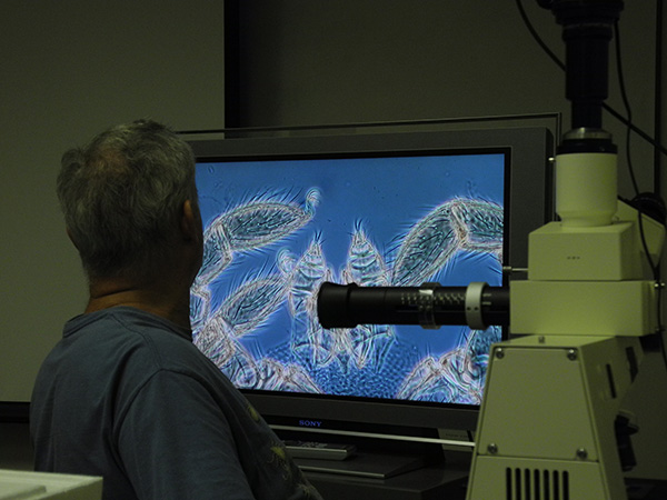
Mite on TV as used for teaching

Mite on TV

With enough technology, mites are coming out of the deep and dark, and into the light, where they of course belong.  We are slowly generating more high quality images and making them available through the collection database.  Check it out, the small can be beautiful.

About the Author: Dr. Hans Klompen is professor in the Department of Evolution, Ecology and Organismal Biology and Director of the Ohio State University Acarology Collection.


Photo follow-up: Diversity in brooding sea anemones


In the last post, I wrote about brooding sea anemones. In this gallery I’ll cover some of the diversity in brooding species and show the various ways in which offspring are brooded. The following are images of animals from the field and of preserved specimens. Click or tap any of them for larger versions.


Internal:
Some species brood offspring internally. In this case, the young simply develop within the body of the adult. Internally brooding animals usually aren’t readily noticeable as such. However, under certain conditions one can sometimes see offspring within the adult.


External:
External brooding is usually more conspicuous in the field than internal brooding. In this behavior, developing offspring are held upon the adult’s body. There may be specializations in the structures of the adult that increase retention and/or protection of the offspring. In other cases, the young may simply be attached to the adult as they would any other surface. Epiactis prolifera offers an example of the simplest case.


In other cases, the offspring are held in modifications of the adult’s body wall. These modifications appear in three general forms:

Pits are depressions in the body wall which hold individual offspring. They can be all over the adult’s column or restricted to certain areas.


Grooves surround the adult somewhat like a moat around a castle.  A groove may be open or closed and is formed by a circular depression in the adult’s body wall.


Chambers are one of the most unusual and interesting structures that sea anemones use to protect externally developed offspring.  They are pouches that go deep into the adult’s body and house many offspring simultaneously. Unfortunately, due to their rarity (only two or possibly three species produce chambers) and Antarctic distribution I have never seen a sea anemone with chambers, so a drawing made from a previously published figure (Carlgren 1901) will have to suffice.

Two brood chambers (holding many offspring) are visible towards the top of this cut-away view of Halianthella kerguelensis.  The one on the left is intact. The one on the right is bisected.

Two brood chambers (holding many offspring) are visible towards the top of this cut-away view of Halianthella kerguelensis. The one on the left is intact. The one on the right is bisected.


Reference:

Carlgren, O., 1901. Die Brutpflege der Actiniarien. Biol. Centralbl., Bd. XXI. Leipzig 468–484.


About the Author: Paul Larson is a PhD candidate at the Department of Evolution, Ecology and Organismal Biology at The Ohio State University studying evolution of marine invertebrates.
email: larson.309@osu.edu
twitter: @mar_inv

Timing is everything: where and when to collect specimens


It’s often said that museum specimens offer us a snapshot into the past. More than simply being a record of what existed where and when, however, specimens can tell us about parts of the life histories of organisms that can’t easily be observed in the long term. As an adult human, I don’t change very much over the course of the year (holidays excluded), but many species undergo seasonal cycles that can be completely missed when collection only occurs during the “field season” (usually summer).

In the course of my work on sea anemones, I came upon a species identity crisis. A group of sea anemones from Alaska were combined with a Japanese species by certain authors, while others considered them to belong to a species found in California. They surely don’t all belong to one and the same group because the Japanese and Californian species have very different reproductive behaviors. In Japan, baby sea anemones are brooded upon the surface of the adult, and in California, the babies are brooded within the adult’s body.

Do the Alaskan sea anemones belong to either the Japanese or Californian species?

Do the Alaskan sea anemones belong to the Japanese or Californian species? -or neither?

The obvious question is “Do the Alaskan sea anemones brood (and, if so, how)?” To figure this out I contacted curators and searched databases from museums around the country and borrowed each specimen from Alaska that I could find (less than 10 in total, and none of them handled by the original author of that species). The specimens all had two important things in common: None had any sign of babies, either in or on their bodies, and they were all collected in the summer. The Japanese species broods primarily in the winter so, if the Alaskan ones are also external brooders, maybe they have just not been collected during the brooding season!

Thanks to a grant from the American Museum of Natural History (AMNH), I began planning the logistics of a field trip to Alaska. The timing for this trip was critical. Too early in the year, and I risk being stuck in Anchorage due to hideous weather at my sampling localities; too late, and I get nicer weather but possibly miss the brooding season. I planned the trip for early April, hoping to hit a sweet spot between the two extremes.

Brooded babies on the surface of Epiactis japonica

Brooded babies on the surface of Epiactis japonica, from Hokkaido, Japan.

My first sampling site was Popof Island. Unfortunately the prime rocky locality (I had identified it in satellite images) for my species of interest was inaccessible due to winter conditions, and most of the rest of the island was sandy shores. It’s also the original locality for the Alaskan species, so that site was a disappointment for my immediate purposes. So far, off to a bad start.

My next sampling site was Adak Island where I had better luck. Not only did I find the species I was looking for, but they were ringed with little baby sea anemones! Before returning home to the OSU Museum of Biological Diversity, I had another equally successful collecting stop on Kodiak Island where I found more of the external brooders. These animals partially answered the question I traveled all that way to investigate: The Alaskan sea anemones were not the same species as the Californian ones (and the Californian ones would need a new name). But are the Alaskan and Japanese species the same, or do they just behave in a similar way?

Epiactis ritteri with a ring of brooded babies (beige blobs)

Epiactis ritteri with a ring of brooded babies (beige). Adak Island, Alaska.

A walk on a nice sandy beach before collecting in the rocky intertidal  on Adak Island

A long walk on a sandy beach on the way to collecting in the rocky intertidal on Adak Island.

Based on anatomical study of the specimens I had already borrowed (and previous collections I’d made of the Japanese species) I had strong reasons to consider that all three groups were different species. Seeing the animals alive in the field gave me even more features to draw from (their color patterns and their resting posture -things which are frequently lost in preserved specimens). By chance I subsequently came across another specimen of the Alaskan species from another museum. This one had been identified by the original author (several years after he published the species description) and also had very large offspring all over its body!

It turns out all the information I needed already existed in previously collected specimens. Was my trip a waste?  Absolutely not! The new collections represent a time between the non-brooding summer season and the late-stage brooding of this new (to me) specimen. It turns out that during this intermediate time, the relatively small offspring are kept within a special groove that forms by a fold in the adult’s body wall and seals the babies inside. As the babies grow, the groove opens up and they simply live on the surface of the adult. My collections are the first record of this type of sealed groove, and they include life stages from early embryos to juvenile sea anemones, and of course the brooding adults. Furthermore, I needed fresh material for DNA analysis (formalin-fixed specimens are extremely difficult to get good DNA sequences from).

The sealed brood groove (horizontal band) in  Epiactis ritteri has partially broken open to reveal an offspring held within it (see two tentacle tips)

The sealed brood groove (horizontal band) in Epiactis ritteri has partially broken open to reveal one of the offspring held within it (see two tentacle tips).

Since the OSU Museum of Biological Diversity doesn’t house a sea anemone collection, I returned the favor to AMNH and donated the new specimens to its collections. The specimens I collected are just as much of a static ‘snapshot’ in time and space as any other single collection event, but when you combine snapshots from different times, you can begin to make a flip-book (or a .gif, if you must) that tells a dynamic story.


About the Author: Paul Larson is a PhD candidate at the Department of Evolution, Ecology and Organismal Biology at The Ohio State University studying evolution of marine invertebrates. email: larson.309@osu.edu twitter: @mar_inv

Museum specimens going online


Skipper butterflies (Erynnis martiallis) are some of the specimens being digitized and imaged at the Triplehorn Insect Collection

Skipper butterflies (Erynnis martiallis) are some of the specimens being digitized and imaged at the Triplehorn Insect Collection

We open the doors to our collections once a year for the Museum Open House and thousands of people from all over the state and beyond stream through our building to marvel over our specimens. Many of them express interest in re-visiting soon.

How amazing would it be to allow people access to our specimens every day at any time? With easy access to the World Wide Web it is possible and natural history museums are digitizing their collections and making their specimens freely available online.

Digitization of plant specimens in the OSU Herbarium.

Digitization of plant specimens in the OSU Herbarium.

Curatorial staff take high quality, ideally 3-D images of each specimen, add metadata and upload them to an online database. This process is labor- and time-intensive, but well worth the effort.

You can read about Museum Specimens Find(ing) New Life Online in this recent New York Times article (10/20/2105.)  And please stay tuned to learn more about specific digitizing efforts going on right here in the collections housed at the OSU Museum of Biological Diversity.


About the authorsAngelika Nelson (Borror Lab of Bioacoustics) & Luciana Musetti (Triplehorn Insect Collection) collaborated on this post.

Going where the wasps are.


On the previous post we talked a bit about visits to the Museum of Biological Diversity and more specifically to the Triplehorn Insect Collection. Today I want to turn around and show you what we see & do when we put our ‘research scientist‘ hats and go visit other collections.

There’s no walk-ins when it comes to visiting research collections. Setting up an appointment with the curator or collection manager is a must. That allows the staff to prepare for our visit, to set up work stations for us, to review the material we are interested in, and to do curatorial work ahead of our visit if necessary.

Collections welcome research visitors because that fulfill their mission of providing service to the scientific community. In return, visiting scientists add value to the collection by providing expert identification to specimens in the collection, and many times helping out with curation and organization of the collection.

When we get to a museum or collection, the first thing we see is, of course, the door. While most public museums have imposing entrances, many times the access to very important research collections is a modest door on the side of a building. The size and type of door absolutely does not reflect the quality of the collections inside.


Once inside, we have access to the inner sanctum of the collections: rows of cabinets filled with drawers filled with dry specimens carefully separated by group; vaults containing insect specimens preserved in ethanol, waiting to be sorted to family, genus, species. And that’s when our work begins!


During a research visit we usually: 1) examine (lots and lots of) specimens, dry or wet, under the microscope, 2) add identification labels to specimens that we recognize, 3) database the specimen label data, and 4) take photos of specimens (and specimen labels). Sometimes we do only 1 and 2, other times we do mostly 3 and 4. It depends on the collection and what we are hoping to accomplish during our visit.


Over the years we had the opportunity to visit many (many!) amazing research collections in various countries. Besides the collections, their rich treasure of specimens, and their dedicated curatorial staff, we also learnt a lot about the places and the people who live there. Looking forward to our next research visit to a collection!


About the Author: Dr. Luciana Musetti is an entomologist and Curator of the C. A. Triplehorn Insect Collection.

Up close and personal: insects and molluscs


Here’s one question I get frequently from visitors: “Why, oh, why, isn’t the Museum of Biological Diversity open to everyone every day?” That’s a very good question! Here’s an answer. Unlike institutions such as the Smithsonian Museum of Natural History, the Field Museum, the Carnegie Museum, or our neighbor the Cleveland Museum among others, our museum largely grew out of a background of higher education and research.  We have a different structure and a different mission than these other very fine institutions.  The most visible outcome of these differences is the fact that we don’t have large display areas and exhibits. We also do not have staff dedicated to public outreach. But it’s good to keep in mind that the MBD collections vary in the kinds of services they provide to the community. Each is unique in it’s own way.

My little corner of the MBD is the Triplehorn Insect Collection. We are a research facility and most of our specimens are only accessible to professional scientists and scientists in training (graduate students, postdoctoral associates, etc.)  This policy gets me in trouble with a lot of people who love insects and would like to come in to “see” (many times that means “touch”) the collection.  So, before anyone else gets hot under the collar about that, let’s try to understand what that policy means.

Dried insect specimens are as fragile as they are colorful and beautiful. The more they are handled and exposed to light and humidity, the faster and more likely they are to get damaged.  The insect specimens in the Triplehorn collection are the result of more than 100 years of careful collecting and curation, many of them were collected in forests and meadows and prairies that do not exist anymore. These specimens are, literally, irreplaceable, and it is our responsibility to keep them intact for many more long years.

Aquatic beetles.

Aquatic beetles. Part of the holdings of the Triplehorn Insect Collection.


Because of that, we restrict access to the specimens to only the people who must use them for scientific study, professionals who have lots of experience with museum specimens and therefore are less likely to damage our precious charges. As the curator of the collection, it is my responsibility to protect and preserve the specimens for the long run. To do that I have to enforce the “restricted access” policy.

Now, the fact that we are a research collection does not mean we don’t welcome visitors.  Quite the contrary! We are committed to sharing our knowledge and love of biodiversity with everyone interested.  While we don’t have exhibits per se, we frequently and happily provide tours of the collection to people from the local community. Or even not so local: our audience is wide and varied, from k-12 to university classes, to family or neighborhood groups, to homeschool groups, to citizen scientists and individuals interested in local and global insect diversity.


Up to now we have been scheduling visits as requests come in and our time allows, but starting this month we in the insect collection will be teaming up with our colleagues in the Mollusc Division of the MBD to offer guided tours of the two collections to the general public on set dates.  This initiative comes as a response to the increased interest in the collections, demonstrated by the increase in visit requests.

Tours will still be arranged in advance, but by specifying which days are open for tours we hope to make the whole process a bit easier and more predictable. The set dates might not work for all visitors, but by working together and establishing a structure for tour activities, we hope to continue serving the community without drastically increasing the work-load of our already overworked staff.


The next available dates for insect collection/mollusc collection joint tours are Friday, October 23rd and Friday, November 6th, from 1pm to 4pm. Total estimated tour time for the two collections is between 45 min to 1 hour/group. Group size limit is 20 adults.  For more information or to schedule a guided tour, please contact Tom Watters or Luciana Musetti.


About the Author: Dr. Luciana Musetti is an entomologist and Curator of the C.A. Triplehorn Insect Collection.

A classy group of land snails


In addition to freshwater molluscs, we also work with terrestrial snails. Our research is concentrated on the Caribbean snail family Annulariidae (ca. 700 species). A few examples are shown below.

About the author: Dr. G. Thomas Watters is the Curator of Molluscs

Hawai’ian Tree Snails – an old and unlikely Ohio connection


Hawai’i is (or was) home to a great diversity of tree snails in the families Achatinellidae and Amastridae. It seemed as if every valley on every island had its own suite of species. Many were quite common. But that was then. Now they have been decimated by the introduction, accidental or otherwise, of invasive hogs and other animals brought by European settlers. Some species, even an entire genus, have become extinct.

Wesley Newcomb

Wesley Newcomb

Wesley Newcomb (1808-1892) was a physician, social activist, and conchologist. Born in New York, he moved from Albany to California in 1849, then to Hawai’i in 1850 due, in part, to his wife’s ill health. There he practiced medicine, served on the Board of Health, became active in the Hawaiian Temperance Movement, and collected a lot of shells. In 1855 he returned to Albany. His collection was purchased by Ezra Cornell, founder of Cornell University, for $15,000, and it resides there still. An avid shell collector, Newcomb traveled to Europe, the West Indies, and Central and South America. He described over 100 species, including many Hawai’ian Achatinellidae and Amastridae.

The Division of Molluscs has a modest collection of Hawai’ian Achatinellidae and Amastridae originating from Newcomb. The collection was either purchased by or traded with Henry Moores in the mid-1800’s. (Henry Moores, 1812-1896, assembled one of the most diverse shell collections of his time. The Ohio State University purchased this collection, 3,500 specimens for $1,750, about 1890). Card stock used for labels cut from postcards date from the 1850s. The collection has an accompanying list of specimens, some notes, and a short letter to Moores. The curious, printed handwriting matches that of Newcomb’s labels now at the Paleontological Research Institute at Ithaca, New York, and there is no doubt that they are Newcomb’s specimens. Specimens have as many as six labels, with different numbers, in the same vials. However, according to Newcomb’s accompanying list, he inserted “card” labels with a number that matched the number on the list. These card labels, often small squares, have numbers written by Newcomb’s hand and can easily be discerned from the later labels added to the specimens. Some specimens are numbered in ink or pencil, but these numbers were added by Moores. The specimens were apparently sent to Moores after Newcomb’s return to the mainland.

Portion of letter to Moores

Portion of letter to Moores

Some aspects of the collection are interesting from a personal view of Newcomb. Many of the specimens were dirty and bear on the small card labels the advice “wash them” (we have carefully done so in a sonic cleaner). The list arrived before the shells as Newcomb tells Moores to “Wait for the Waggon! (Express)” And one label bears the opinionated observation: “‘guernea‘ W.H.P. [crossed out, then added:] A. perversa? Swains. ‘guernea‘ of some fool.” Newcomb’s “fool” was apparently contemporary fellow Hawai’ian conchologist, William Harper Pease.


 Below are some of Newcomb’s own specimens now in the Division of Molluscs.

About the Author: Dr. G. Thomas Watters is the Curator of Molluscs.

Remembering Carol B. Stein — Tetrapod Collection


Carol B. Stein, an Assistant Curator for the Natural History Department at the Ohio State Museum passed away this past March 2015 on 6 December 2010. Though her primary focus was in the Molluscs, she provided over 400 specimens to the Tetrapod Collection. In remembrance of all the collecting she did for the museum, we dedicate this post to her and her contribution to the Tetrapod Collection.


About the Author: Stephanie Malinich is the Collection Manager for the Tetrapod Collection.


Preparing the Dead – the Dying Art of Preparing Mammals and Birds


Looking at the mammal and bird skins in the OSU Tetrapod Collection, visitors commonly have some questions:

“Where did the specimen come from?”

“What did you do with the insides of the specimen?”

“Why are the specimens’ eyes white?”


To properly answer these questions, we need to go back in time to when naturalists, such as Charles Darwin or John James Audubon, went out into the field to collect specimens. When they captured a bird, they would remove the insides of the specimens to ensure that the skin would not spoil and rot before other naturalists could view and study it. Today, we continue to use very similar methods to preserve specimens, as well as new methods such as freeze drying. – Now going back to the visitors’ questions:

“Where do the specimens come from?”

Trays of brown and white waterfowl laying out on trays to be reorganzied.

Trays of prepared waterfowl specimens laid out to be reorganized.

In the past collecting birds and mammals was a very active process, very similar to today’s trophy hunting. Collectors would go out with shotguns, mist nest and traps to catch animals and collect them. Today, the most common way of getting specimens for collections is by salvaging already deceased animals. To do this, our collection possesses special permits (known as salvage permits) allowing us to salvage birds and mammals from all over the state of Ohio.

“What do you do with the insides of the specimen?”

The tendons found inside the tail of a North American Beaver.

The tendons found inside the tail of a North American Beaver.

We remove most of the bones and tissues of specimens during preparation, and the carcass is used in two ways: Most carcasses we examine internally to determine the sex of the specimen and to look at the animal’s last meal. This will provide some information about the ecology of the animal, what and where it has been feeding. Once that information has been recorded the carcass is disposed of. Rare specimen carcasses are put in a tank of dermestid beetles for a couple weeks till only the bones remain. Once cleaned, the bones are added into the collection for future research use.

“Why are the specimens’ eyes white?”

A row of Rose-breasted Grosbeaks males.

A row of Rose-breasted Grosbeaks males.

To ensure that our specimens will last 500 years or more we remove as much of the organic material as possible. This means that we remove the eyes, brain, body and a majority of the muscle tissue. After removing the organic tissues and matter, we fill the now empty skin with white cotton to represent the body 3 dimensionally. Since the eyeballs have been removed the white cotton in the inside of the head is visible through the eye sockets – hence the white eyes. By removing most of the organic material, we reduce the potential for attracting museum pests, such as the dermestid beetle also known as the flesh-eating beetle – you can imagine what this beetle would do to a specimen with intact insides.

Today, the traditional method of preparing specimens by skinning is slowly being lost. Newer methods of freeze drying specimens, using large freeze drying machines that pull all moisture out of the specimen, have become more popular. Freeze drying allows for more organic material to be preserved which in turn provides more material to use for DNA testing of specimens. In our collection, we do a combination of freeze drying and traditional methods of preparing specimens, depending on size and overall condition of the specimen.

How Can You Help?

To add to our collection we need citizen scientist, like yourself, who find a dead bird or mammal, write down the day and place where they found it, and donate it to the collection. On average, we get 10-15 new birds a month, which we put in a freezer to prepare at a later date. These specimens once accessioned will “live” in the collection for a century or longer.

Something to note: without proper permits, a citizen is not allowed to possess a deceased migratory bird, due to the Migratory Bird Act (more info at http://www.fws.gov/laws/lawsdigest/migtrea.html).


About the Author: Stephanie Malinich is the Collection Manager for the Tetrapod Collection.