
Basic Information | Advanced Information | Diagnostic Information
Tomato Diseases | Bacterial Canker
Bacterial Canker
Symptoms
Bacterial canker infections show a wide range of symptoms and can affect plants at all growth stages. Key diagnostic symptoms are provided for each tissue type.
Stem:
- Raised pustules on young seedlings
- Reddish-brown necrotic cankers, especially on older tissue
- Reddish-brown vascular discoloration, especially at base and nodes


Images of bacterial canker on tomato stems (left; center) and a pepper stem (right).
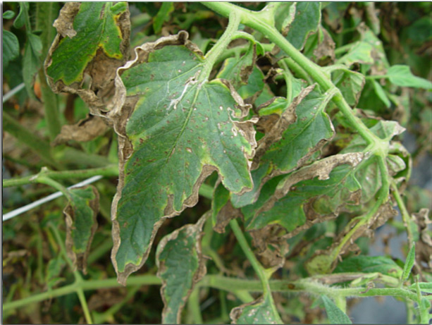
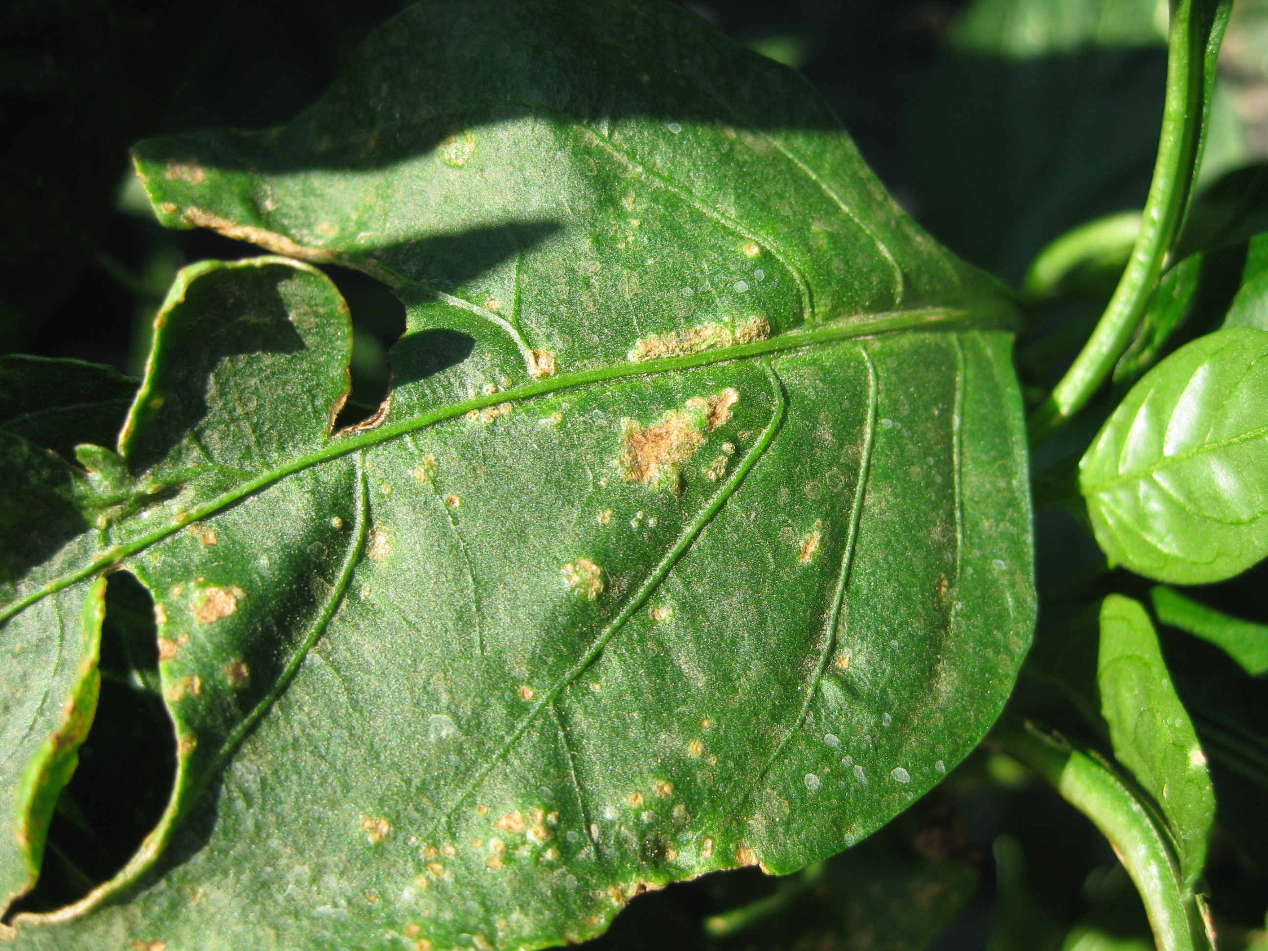
Leaves:
- Raised pustules on young seedlings
- Necrotic marginal leaf tissue adjacent to a thin band of chlorotic tissue (firing)
- On severely infected tissue necrotic lesions with minimal chlorosis may be observed

Roots:
- There are no diagnostic symptoms associated with root tissue
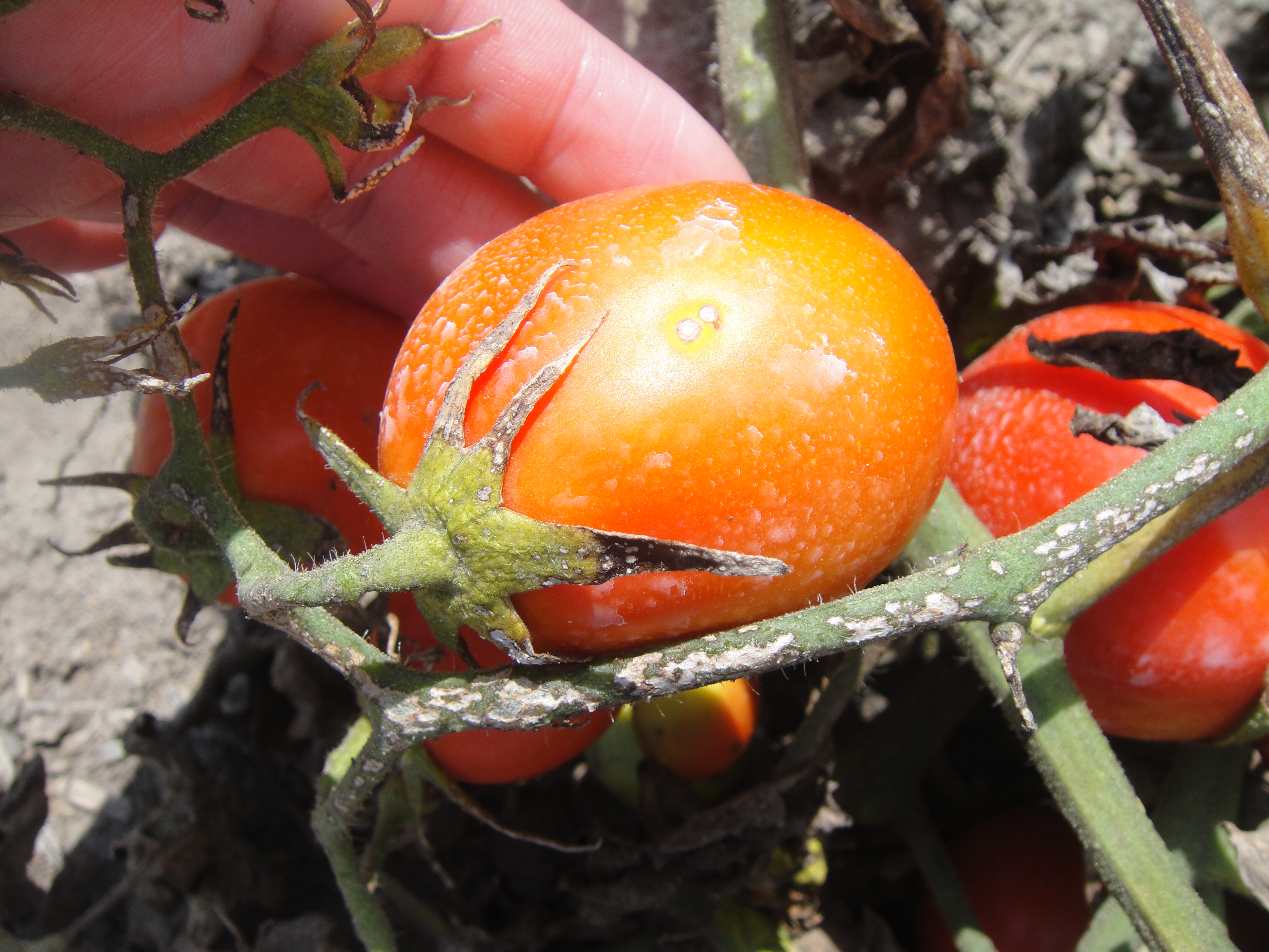
Fruit:
- Field grown tomatoes – small raised tan colored spots surrounded by a white halo on green and red fruit
- Greenhouse grown tomatoes – internal web-like appearance

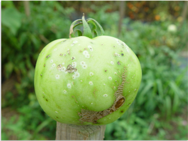

Symptoms of bacterial canker on tomato fruit (left; center) and pepper (right).
Signs
Bacterial streaming from the margin of a leaf, stem or fruit lesion.
Often Confused With
- Bacterial Spot – Look for large crusty lesions on the fruit with no white halo as indicators of bacterial spot.
- Bacterial Speck – Look for small black pin-point lesions on the fruit with no white halo as indicators of bacterial speck.
- Early Blight – Look for lesions with concentric rings and chlorosis as indicators of early blight.
- Mite Damage – Using a dissecting microscope look for live mites on the leaf surface as indicators of mite damage.

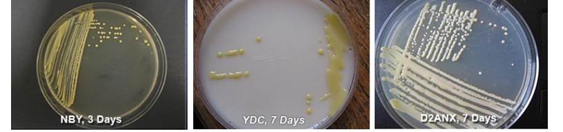
Isolation Media
Nutrient Broth Yeast (NBY) (pdf) Extract medium is a non-selective medium used for subculturing suspected clavibacters. Clavibacter michiganensis subsp. michiganensis colonies are yellow, round, entire and convex after 3-5 days at 82 °F.
Yeast extract-dextrose-calcium carbonate (YDC) (pdf) medium is a non-selective medium used for subculturing suspected xanthomonads and clavibacters. Xanthomonas spp. colonies on YDC medium are yellow, mucoid, round, entire and raised after 3 days at 82 °F.
D2ANX medium (pdf) is a semi-selective medium. Colonies grow slowly on D2ANX medium. After 7-10 days at 82 °F, colonies are light-creamy yellow, mucoid, round, entire and raised.
Available Rapid Diagnostic Tests
- Agdia ImmunoStrip®
- Agdia ® Enzyme Linked Immunosorbent Assay (ELISA)
- Envirologix™ DNAble Quick Stix
- Conventional Polymerase Chain Reaction (PCR) assays
- primers CMM5/6 CMM5/6 (pdf)
- primers CMM3/4 CMM3/4 primers (pdf)
- primers PSA8/R PSA8/R (pdf), PSA primers (pdf)
- Quantitative real time PCR Assay
- primers RZ ptssk protocol 10/11, probeRZ-ptssk12 PTSSK primers (pdf)