What Is It? | Facts in Depth | For the Professional Diagnostician
Tomato Diseases | Tomato Pith Necrosis Fact Sheets
Tomato Pith Necrosis
Symptoms
Foliar symptoms of pith necrosis are similar to early bacterial canker disease symptoms (wilting and marginal leaf necrosis); therefore internal stem symptoms should be used to distinguish between the two diseases.
Stem
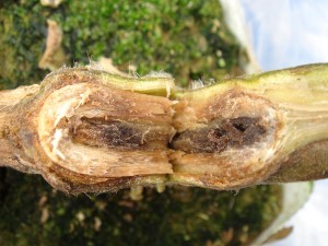
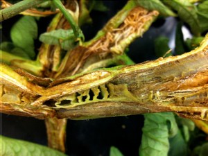
- When stems are cut longitudinally, the pith can be severely degraded or hollow, dark brown to black in color and/or have a ladder-like appearance. Early symptoms are browning of the pith.
- Stems may be swollen and have a large number of adventitious roots associated with them.


Signs
Bacterial streaming may occur from the margin of a stem lesion using a microscope but many samples may need to be tested before observing bacterial streaming.
Often Confused With
- Bacterial Canker – Look for “bird’s eye” spotting on the fruit. Fruit symptoms with pith necrosis are rare. The pith of stems infected with the bacterial canker pathogen is generally white and spongy.
- Bacterial Wilt – Leaves wilt and plants collapse suddenly. Brownish discoloration begins in vascular tissue but moves into the pith later. Copious amounts of bacteria stream into water from infected stems and can be seen with the naked eye. Similar bacterial streaming not observed for pith necrosis. Bacterial wilt is extremely rare in temperate climates in the US.

Isolation Media
Nutrient Broth Yeast (NBY) extract medium (pdf) is a non-selective medium used for subculturing suspected tomato pith necrosis pathogens. Pseudomonas corrugata and P. mediterranea colonies after 3 days at 82°F are yellow-brown, round with a curled margin, and flat.
Yeast extract-dextrose-calcium carbonate (YDC) medium (pdf) is a semi-selective medium for isolating P. corrugata and P. mediterranea. Colonies of both species on YDC medium are yellow with a green center after 5 days at 82°F. Colonies are mucoid, round, curled and flat. Older colonies are dry and wrinkled.
Pseudomonas F (PF) medium (pdf) is a semi-selective medium for isolating pseudomonads. Colonies of P. corrugata and P. mediterranea are non-fluorescent on PF medium. Colonies of P. cichorri, P. viridiflava, P. marginalis are fluorescent on PF medium. After 5 days at 82°F colonies of all species are creamy, yellow-brown in color, round, curled and flat. Older colonies are dry and wrinkled.

Available Rapid Diagnostic Tests
- Conventional Polymerase Chain Reaction (PCR) Assays
- Primers PC1/1 and PC1/2 (P. corrugata) PC1 primers (pdf)
- Primers PC5/1 and PC5/2 (P. mediterranea) PC5 primers (pdf)
