Posts

Porifera

An Introduction to the Sponges

Many of us may find it easy to appreciate the diversity of animals that inhabit our planet. Whether it be a bird, insect, or mammal, we humans are often drawn towards some sort of fascination of their mere existence. But what preceded all of the animals we see around us? What may come to mind are images of dinosaur, trilobite or coral fossils, but there existed animals much less complex than any of these. Porifera, or sponges, represent some of the most primeval of animals, lacking body symmetry or specialized organs.  Instead, their body consists of specialized, individual cells that serve different functions for these filter-feeding, sedentary organisms (Blair, 2009). Sponges can be found worldwide, from shallow reefs to deep ocean trenches. They inhabit both marine and freshwater environments, and come in a variety of shapes and sizes. If these organisms represent such ‘ancient’ animals, how old are they? The oldest reliable sponge fossils date back 535 million years ago from Northern Iran (Antcliffe et al, 2014). In addition, sponges are thought to have diverged from the animal phylogeny during the Precambrian, which lasted up until 540 million years ago (Antcliffe et al, 2014). Their basal status in Metazoa, or animals, and ancient lineage represent just a portion of the significance of these bizarre organisms.


Porifera Phylogeny

The group of organisms known as sponges (Porifera) is considered the earliest branching group of Metazoans, or animals, with fossils described from the Vendian Period, dating back 650-543 million years ago (Porifera: Systematics, 2006). Phylogeny in this phylum, or group of organisms, is an ongoing debate, with the current consensus viewing sponges as possibly mono- or paraphyletic (Blair, 2009). Monophyly would indicate a recent common ancestor of all sponges, while paraphyly would indicate that the group of organisms we regard as sponges is actually made up of groups that developed separately over a relatively extensive time. The sponge phylum consists of four currently recognized classes; the Hexactinellidae, Demospongiae, Calcarea and Homoscleromorpha. The relationship between these four classes is still unresolved (Wörheide et al, 2012). Gross morphology suggests the clade is monophyletic.  With the advent of molecular systematics, this monophyly was put into question; however, after extensive sampling and inclusion of specimens from all classes, one recent paper suggests monophyly may be the agreed relationship (Wörheide et al, 2012). Regardless, Hexactinellida and Demospongia are both regarded as being monophyletic, representing those sponges which contain silica-based skeletons (Blair, 2009).

One of the main diagnostic features of sponges had previously been their spicules, which constitute the hard support structure of these organisms. This was later proven to be an inaccurate means of identification, as currently existing sponges have been discovered with solid calcium-based skeletons, matching some features observed in the fossil record (Porifera: Systematics, 2006). Phylogenetic analysis of Porifera is conducted using mitochondrial DNA sequences, in conjunction with analyses of morphological features as well (Wörheide et al, 2012). Porifera are not just significant for their roles in ecology, pharmaceuticals, and commercial products but also in developing hypotheses of what the last common ancestor of all animals could have been.


phylogeny

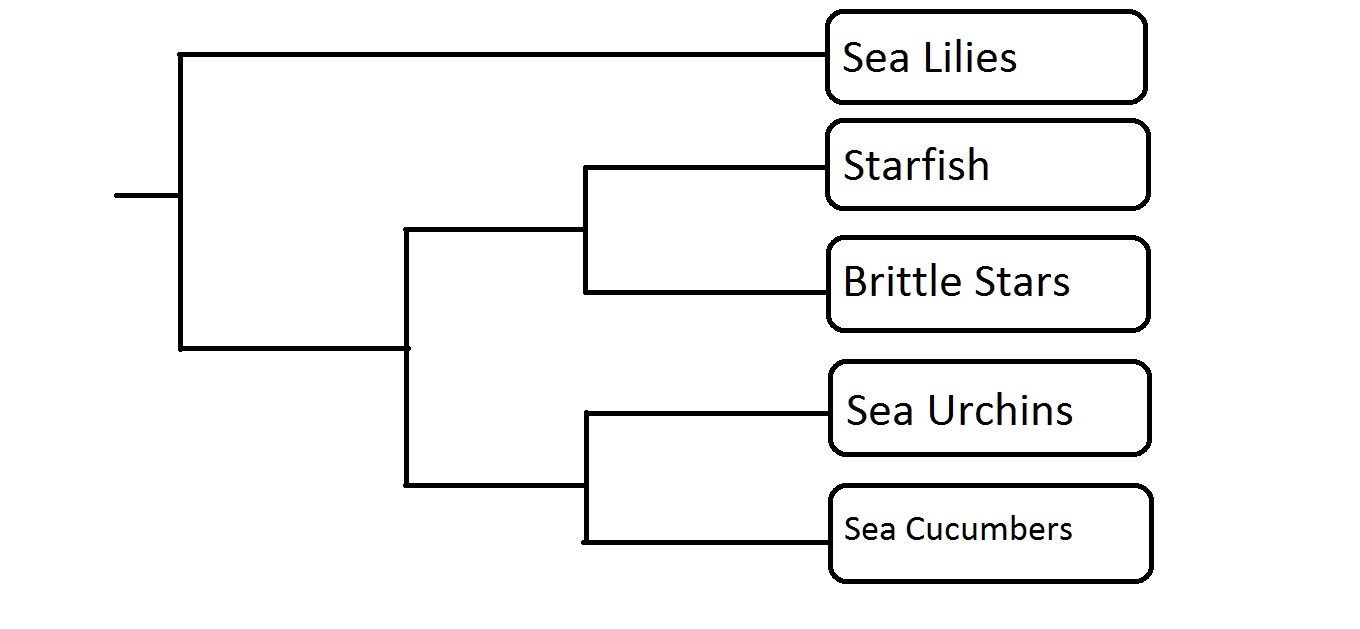
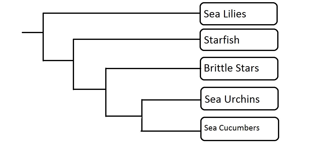
Two proposed models for Porifera phylogeny.  Hex= Hexactinellida, Demo= Demospongia, Cal= Calcarea, and Homo= Homoscleromorpha.  In the left tree, Homoscleromorpha and Calcarea are more closely related to the rest of Metazoa than the other two sponge classes.  In the right tree, all classes share a common ancestor. (Adapted from Wörheide et al, 2012, by Dylan Sedmak)

Fossil Record

          Porifera are the first animals on the metazoan phylogeny, having diverged from choanoflagellates 1020 million years ago (mya). The sponge group Hexactinellida diverged from the Demospongia group around 750 mya and it is estimated that the Calcarea group later diverged from the other two groups an estimated 754 mya (Sperling, et al., 2010). Porifera are the most primitive of animals and thus have an early branch on the animal phylogenetic tree, so they’re likely candidates for Precambrian ancestry (Gehling & Rigby, 1996). To understand this, one needs to look at two sponge groups: the Hexactinellids and the Demosponges. These are the two oldest sponge groups, and both have siliceous spicules. Sperling, et al., (2010) suggests that these spicules must have evolved before the common ancestor of Hexactinellids and Demosponges, which means that these spicules were present in the Precambrian, but not fossilized.

Sponges have a fossil record that extends back further than 500 million years. The oldest fossil found for Hexactinellids are siliceous spicules that were found in Northern Iran and date back to approximately 535 mya and the earliest fossil found for Demosponges came out of Siberia and was dated to be 523.5-525.5 mya (Antcliffe, et al., 2014). These fossil remains appear to be dated around the Precambrian-Cambrian boundary, which is associated with great diversification of animals. Finding fossils for the Calcarea sponges is very difficult because they are not as diverse as the other two groups and they lack siliceous spicules which makes it difficult to find preserved specimens (Antcliffe, et al., 2014).

Finding these fossils and correctly identifying them as sponges is a difficult task, as most reported sponge fossils tend to be volcanic shards or inorganic crystals (Gehling & Rigby, 1996). One paper by Antcliffe, et al., (2014) discusses 20 potential candidates for being the oldest Porifera fossil found. This paper also discussed how it can be difficult to define the criteria that determines the oldest fossil because there is no substantial studies done on the formation of cells that make up sponges. There are no studies that study fossilized sponge spicules that look like today’s sponges or vise versa – looking at potential spicules that look nothing like spicules we find today. Antcliffe, et al. (2014) also made the point that these potential fossils may be misinterpreted as individuals rather than part of a larger organism, which further complicates the fossil record.

Finding just the fossilized spicules makes it difficult for scientists to get a clear picture of the shape and form these sponges took. Sponge fossils found in Australia that date back to the Ediacaran period (Precambrian) give some insight into what these first sponges must have looked like. Gehling and Rigby (1996) found that these fossils were formed in hypo-relief on sandstones, with external molds of fossils commonly found. These fossils indicate that the sponges were dome shaped with an osculum at the apex. Since there are no siliceous spicules that have been found before the Precambrian-Cambrian boundary, Gehling and Rigby (1996) note that the siliceous spicules would not be expected to survive longer than the organism’s tissue during fossilization due to being preserved in sandstone substrate.


Molecular Clock

        The molecular clock for Porifera suggests that their origin was prior to the Cambrian explosion. This fits with the assumptions made by Sperling, et al. (2010) that based on the siliceous spicule fossils of Hexactinellids and Demosponges, these spicules must have evolved prior to the Cambrian explosion. Though this indicates a gap in the fossil record, molecular clock analyses can still be done to determine divergence time estimates.

          A paper by Antcliffe, et al., (2014) discussed how it is sometimes difficult to get accurate divergence times because fossils are needed to help the molecular clock to be more accurate in its predictions. The dates of the fossils help the clock analyses to better date divergence times. These dates were found using a small number of nuclear housekeeping genes, which are genes that are generally used to date animal phylogenies. This paper found that the housekeeping genes indicated Porifera diverged from the animal lineage around 800 mya in the Precambrian.

Another paper by Sperling, et al. (2010) did two sets of molecular analyses. One set was with multiple sponges from Hexactinellida and Demosponges. The second set was done without Hexactinellida because the Hexactinellid group is the first group that diverged from the metazoan lineage. Their analyses led to the conclusion that the Silicea (both Hexactinellids and Demosponges) originated around 759 mya and also Demosponges greatly diverged within its own clade around 699 mya. For more divergence dates in the Poriferan phylogeny, refer to Figure 2.

phylogeny tree porifera

Figure 2. This timeline represents the estimated divergence times of the Porifera clade. Data represented in this timeline comes from class notes, Sperling, et al. (2010) and Antcliffe et al. (2014). Porifera diverged from the animal (Eumetazoan) lineage approx. 800 mya with Demosponges and Hexactinellids diverging 759 mya and Calcareans diverging 754 mya. Demosponges further diversified into its own clade 699 mya and Calcareans also further diversified 488 mya. Hex. = Hexactinellids, Demo. = Demosponges, Cal. = Calcareans. Phylogeny created by Sarah Petersen.


Key evolutionary innovations

Slime

Some sponges are able to produce slime as a defense against debris or other marine organisms. The amount of slime a sponge can make depends on the type of sponge. There are sponges that have absolutely no slime at all, while others only produce a little and others can produce a lot (Ackers, Moss, & Picton, 1992). The slime certain sponges produce is actually toxic. This natural defense comes from metabolic waste produced by the actual sponge or from toxins that the sponge has modified from these original chemicals (Goudie, Norman, & Finn, 2013).

slime

Figure 3. In this image, a student is seen displaying the sponge’s natural slime excretions in a laboratory setting. Picture taken by Natalie Iannelli.
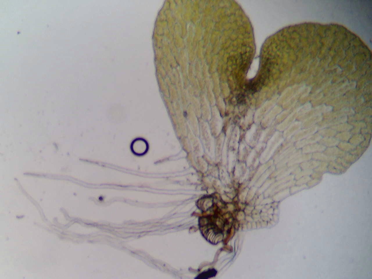
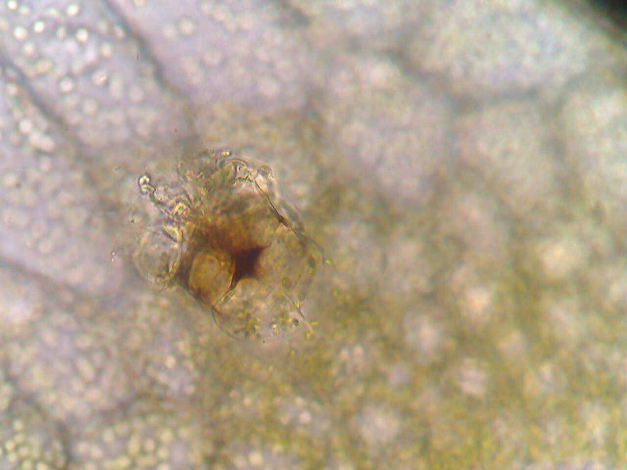
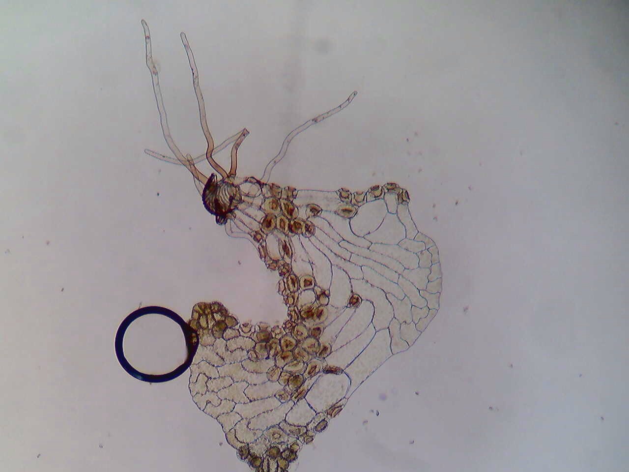
 

Spicules

Spicules are part of the sponge’s “skeleton” and help to give it shape. There are a wide variety of spicules that can be seen in varying sponges. They can help us determine when different sponge species evolved because of their ability to be genetically determined. The environment can also cause spicules to develop in different shapes or sizes and for more than one type of spicule to be present at a time. Spicules are thought to help sponge in a variety of other ways, such as by helping sponge larvae maintain buoyancy, allowing the larvae to reach a spot to settle, enhancing reproductive success, and catching prey (Uriz, Turon, Becerro, & Agell, 2003).

microscope spicules

Figure 4. A microscopic view of a sponge slurry; the spicules can be observed. The view is on a compound microscope at 400X magnification. Picture taken by Christine Koporc and Sarah Petersen.


Toxins

Sponges are able to reuse toxins from other organisms around them, though they can also create their own toxins or in collaboration with the microbes that live inside of them. Many sponges have been found to release highly toxic chemicals and these excretions make up some of the most toxic chemicals in nature. Many of these toxins are used to protect themselves against predators or to outcompete other organisms in a crowded area, but they can be used by humans as well. It has been determined that some of these chemicals could be used in anti-cancer, anti-malaria, and pain control applications (Queensland Museum, 2012).

Apoptosis

Cell death, or apoptosis, is when a cell determines that it is no longer needed and it uses an intracellular death program to get rid of the excess cells. This is a common occurrence in organisms and it even takes place in healthy humans. For example, in a normal healthy human, billions of bone marrow and intestine cells die every hour. There are various reasons for this phenomenon, some of which are in order to properly form a structure when an organism is an embryo or to help ensure that the number of cells does not become too large (Alberts, et al., 2002). Apoptosis first developed in the transition between sponges and their ancestor, meaning that sponges were the first organisms to have a trait of this sort (Werner & Muller, 2003).

Water flow

Sponges contain holes in their bodies to maximize efficiency of water flow. The more surface area there is to absorb nutrients it gets from the water, the better off the sponge will be. The sponges have porocytes on the outside which are openings the water flows into. It then flows out through an opening called the osculum. They are able to pump the water because of flagella on the inside of their cell walls (Porifera: Systematics, 2006).

water flow poriferaFigure 5. This diagram illustrates the method sponges use in order to create water flow through their bodies. Image created by Christine Koporc.

 Video 1. This video demonstrates the water flow system in a sponge. A neon green dye was injected into the sponge and the dye can be seen coming out of the sponge on the other side. Video taken by Natalie Iannelli, edited by Christine Koporc.

Tissue Regeneration   

Sponges have the ability to regenerate their tissue. A study of the capacity of sponges to redevelop conducted at the Carmabi Marine Research Institute located in the Caribbean showed that there are three phases as to how this happens. The first phase is where the damaged surface is closed off by a scar like tissue. During phase two, the tissue changed back to the normal appearance of the surface of the sponge. The only difference is that there is a depression in the surface.The third phase is the filling of the depression. The regeneration of the sponge does depend on the species; some sponges regenerate faster than others. The ability of sponges to regenerate is an important evolutionary characteristic to their survival because they are the food source in reefs for many fish species as well as turtles (Hoppe, 1988).

tissue regeneration porifera

Figure 6. The result of a sponge slurry regenerating. The red masses that can be seen are what has formed after a couple days since the sponge was broken down in a blender. Picture taken by Christine Koporc.

Immune System

Studying the immune response of sponges has peaked an interest in the medical community as antibiotic resistance has become more of a problem. Sponges filter a lot of water during their lifetime. That water is not only composed of the food they need to survive, but also numerous amounts of viruses, fungi, and bacteria. On the surface of the sponge there are special receptors called lipopolysaccharide or LPS which is a protein that allows them to detect bacterial endotoxins. The sponge has the capability to detect what kind material it is filtering through physical and chemical means. It also is able to rid itself of these unwanted pathogens on a molecular basis. It has what is called a LPS-interacting protein and a macrophage-expressed protein that are activated depending on what its receptors recognize. It was discovered by a man named Metchnikoff that sponges use phagocytosis to kill off bacteria as well. Phagocytosis is the ingestion of bacteria or other kinds of material by a cell. Using its detection methods and the way it kills bacteria, viruses, and fungi, the sponge is able to eliminate the unwanted organic material to keep it from dying (Wiens et al., 2005)


Sources:

Ackers, R. G., Moss, D., & Picton, B. E. (1992). Sponges of the British Isles (“Sponge V”) (p. 7).

Alberts, B., Johnson, A., Lewis, J., Raff, M., Roberts, K., & Walter, P. (2002). Programmed Cell Death (Apoptosis). Molecular Biology of the Cell.

Antcliffe, J.B., Callow, R.H.T., & Brasier, M.D. (2014). Giving the earliest fossil  record of sponges a squeeze. Biological Reviews, 89, 972-1004.

Blair, J. E. (2009). Animals (Metazoa). In S. B. Hedges & S. Kumar (Eds.), The Timetree of Life (p. 223). Oxford University Press.

Gehling, J.G., & Rigby, J.K. (1996) Long expected sponges from the Neoproterozoic Ediacarda fauna of south Australia. Paleontological Society, 70(2), 185-195.

Goudie, L., Norman, M. D., & Finn, J. (2013). Sponges: A Museum Victoria Field Guide (p. 18).

Hoppe, W. F. (1988). Reproductive patterns in three species of large coral reef sponges. Coral Reefs, 7, 45–50. doi:10.1007/BF00301981

Porifera: Systematics. (2006). Retrieved February 06, 2015, from http://www.ucmp.berkeley.edu/porifera/poriferasy.html

Queensland Museum. (2012). Toxic Sponges & Pharmaceutical Properties. Retrieved October 02, 2015, from http://www.qm.qld.gov.au/Find+out+about/Animals+of+Queensland/Sea+Life/Sponges/Toxic+sponges+and+pharmaceutical+properties#.VNt_EObF-gu

Sperling, E.A., Robinson, J.M., Pisani, D., & Peterson, K.J. (2010) Where’s the glass? Biomarkers, molecular clocks, and microRNAs suggest a 200-myr missing Precambrian fossil record of siliceous sponge spicules. Geobiology, 8, 24-36.

Uriz, M.-J., Turon, X., Becerro, M. A., & Agell, G. (2003). Siliceous spicules and skeleton frameworks in sponges: Origin, diversity, ultrascrutural patterns, and biological functions. Microscopy Research and Technique, 62(4), 279–299.

Wiens, M., Korzhev, M., Krasko, A., Thakur, N. L., Perović-Ottstadt, S., Breter, H. J., … Müller, W. E. G. (2005). Innate immune defense of the sponge Suberites domuncula against bacteria involves a MyD88-dependent signaling pathway: Induction of a perforin-like molecule. Journal of Biological Chemistry, 280(30), 27949–27959. doi:10.1074/jbc.M504049200

Werner, E., & Muller, G. (2003). The Origin of Metazoan Complexity: Porifera as Integrated Animals. Integrative & Comparative Biology, 43(1), 3–10.

Wörheide, M., Erpenbeck, D., Larroux, D., Maldonado, C.,  Voigt, M. , C. B. and D. V. L. (2012). Deep Phylogeny and Evolution of Sponges (Phylum Porifera). Advances in Marine Biology, 61.

Ferns

With over a quarter of a million plant species estimated to be living today, 13,000 may seem like a drop in the bucket, but ferns actually show a major evolutionary step for plants as we know them today (Pearson Education, 2015). Ferns contain a variety of different types of plants under the supergroup archaeoplastida. Although ferns do not have an official classification; they are a part of the subkingdom embryophyta, which contains all land plants. 

There are two different classifications of ferns: the monilophytes and the pteridophytes. Monilophytes include true ferns like the leptosporangiates, the largest group of ferns including over nine thousand species worldwide, while the term pteridophytes include both ferns and some other vascular plants. The term “pteridophyte” has fallen out of favor since it is not one monophyletic grouping like the monilophytes (Schuettpelz, 2004).

The main organism that we see, the growing and the adult fern, known as a sporophyte, is diploid. Diploidy in organisms means that they have two sets of chromosomes. Many organisms that we see today like cats, dogs, and people are also diploid organisms.

The History of Ferns

 ferntree
Phylogeny of ferns. The above figure shows the phylogeny of ferns with divergence dates. A phylogeny is like a family tree and shows how different organisms are related (Pryer et al., 2004).

The first fossils with fern characteristics, embryophytes, begin to appear on record during the middle of the Devonian period about 300 million years ago (USDA, 2006). The Devonian period lasted from 416 million years ago (mya) to 360 mya, and is characterized by the radiation and diversification of plant species and ended with a large scale extinction event that scientists believe was due to global cooling and rising sea levels.

These early ferns split off from the green algae around 960 mya. These are some of the earliest land plants known. The “great fern radiation” is when many modern families of ferns first began to appear. This radiation occurred in the late-Cretaceous period (Bhattacharya, 2009).  Today, roughly 13,000 different species make up the class Polypodiopsida (The Plant List, 2010).

During the Carboniferous period, giant ferns dominated the landscape that grew 9 to 12 meters in height. This was before the time of the angiosperms, flowering plants that produced fruits and seeds, so there was little competition for land. There are still some giant ferns that exist today like Angiopteris evecta, or the giant fern, that can grow up to 7 meters tall and is native to Indonesia, New Guinea, coastal northern Australia and the south and west Pacific Islands (Christenhusz , 2011).

What Makes a Fern a Fern?

Ferns are classified by their vascular tissue, spore production, and lack of flowers and seeds. These plants have three major parts: the rhizome, the frond, and the sporangia. Generally, one or more fronds are attached to the rhizomes by a stipe. The rhizomes are a specialized root structure that draws up nutrients from the soil. The frond is the leaf structure of the fern while the stipe is the technical term for the stalk of the fern. The sporangia are the structures that hold the spores necessary for reproduction (University of Waikato, 2010).

Vascular plants can be differentiated from non-vascular plants by the presence of xylem and phloem. The xylem and phloem work like arteries and veins and help transport nutrients throughout the body of the plant. As the first vascular plants, ferns were able to grow taller rather than wider. Non-vascular plants, like mosses, needed to grow close to the ground in moist areas in order to gain all the nutrients needed to support the organism.


Fern Life Cycle. The video link above shows the life cycle of a fern.

Like mosses, ferns reproduce via spores instead of by seeds. These spores are dispersed via wind and water. When the spores land in a suitable habitat they will begin to grow. The spores of the plant are very small and must be viewed under a microscope because they are about 1/10th of a millimeter in size, and because of their almost invisible nature, ferns and other spore producing plants are known as cryptogams. The term cryptogam refers to their hidden reproduction, and literally means “hidden marriage” (Sytsma, 2014).

These spores, as mentioned before, are housed in the sporangium. In many species of ferns, the sporangium is found on the underside of the leaves, or fronds, of the adult fern. These tend to be grouped into sori (singular, sorus) which are visible to the naked eye as little brown spots. In many species of ferns these sori are protected by an indusium which is a thin membrane that protects the underdeveloped spores and sori. When the spores are ready to be dispersed the indusium shrivels away.

IMG_1506
Indusium on spores of a fern. In the image above, the tan parts on top of the darker sporangium, are already starting to shrivel up. This structure is called indusium. Scale: ~ 3 cm.

20150210_142413(1)
Spores from the underside of a fern. Not all ferns have indusium. Individual frond: ~ 12 cm.

The spores are produced in the sporangium via meiosis. Meiosis is a form of cell division that reduces the number of parental chromosomes in half forming four daughter cells. This is the same process that produces eggs and sperm in animal species. The four daughter cells are haploid which means they contain only one set of chromosomes. These haploid spores are then dispersed by wind or water and will germinate if they land in a suitable environment. The spores then grow into a gametophyte which is also a haploid organism.

The gametophyte is a thin, photosynthetic structure that is only one cell layer thick. The gametophyte produces the sex cells: the egg and the sperm. It also contains two different types of sex organs to produce both of the sex cells. The organs are known as the antheridium, which produces sperm, and the archegonium, which produces eggs. The archegonium is a funnel shaped grouping of 4 cells which allows the sperm cell to swim down into it to meet the egg. The sperm have to swim to the eggs, so the gametophytes need to be in  a very moist environment (The Plant List, 2010).

image7(1)
Female gametophyte. The female gametophyte contains archegonium near the crevice at the top of the gametophyte. Scale: ~1 mm. x 1 mm.

Image-1(3)
Detail of archegonium. The archegonium house the female gamete, the eggs. This is what the male gamete, the sperm, travel into for fertilization. Shown at 40x magnification.

Image-1(2)
Male gametophyte. The preserved, male gametophyte has antheridium, shown as the brown spots. These antheridium once contained sperm. Shown at 10x magnification.

Since this is only one small  part of the life cycle, the gametophyte does not have a root system, but instead has rhizoids. These rhizoids are like miniature roots that serve to anchor the gametophyte and gain nutrients. Once the haploid egg and sperm meet they begin to grow into the diploid organism that we are more familiar with.


Movement of antheridia. The video link above shows the movement of the antheridia through a solution. The antheridia are the tumbling sphere shaped organisms moving rapidly across the screen.

A lot of fern characteristics are shared among other land plants. Mosses also reproduce with the use of spores, and different flowering plants have vascular tissue. In total, it is the combination of everything listed that truly makes a fern a fern.

References

Bhattacharya, D., Yoon, H. S., Hedges, S. B., and Hackett, J. D. (2009). “Eukaryotes.” The Time Tree of Life. 116-120. Retrieved from http://timetree.org/pdf/Bhattacharya2009Chap08.pdf

Christenhusz , M. (2011). Angiopteris evecta (giant fern or king fern). Retreived February 29, 2015 from http://www.nhm.ac.uk/nature-online/species-of-the-day/biodiversity/loss-of-habitat/angiopteris-evecta/

Eberle, J. R., and Banks J. A. (1996). “Genetic Interactions Among Sex-Determining Genes in the Fern Ceratopteris Richardii.” Genetics, 142, 973-85. Retrieved from http://www.genetics.org/content/142/3/973.full.pdf

Pearson Education. (2015, January). Estimated Number of Animal and Plant Species on Earth. Retrieved March 3, 2015. Retrieved from http://www.factmonster.com/ipka/A0934288.html

Pryer, K., Schuettpelz, E., Wolf, P., Schneider, H., Smith, A., & Cranfill, R. (2004). Phylogeny and evolution of ferns (monilophytes) with a focus on the early leptosporangiate divergences. American Journal of Botany, 9(10), 1582-1598.

The Plant List. (2010). “Pteridophytes (Ferns and Fern Allies).” The Plant List — A Working List for All Plant Species, 1. Retrieved on 12 Feb. 2015 fromhttp://www.theplantlist.org/browse/P/

United States Department of Agriculture. (2006, March 5).  Fern Questions and Answers. Retrieved March. 5, 2015 from http://www.usna.usda.gov/Gardens/faqs/fernsfaq2.html

Sytsma, K. (2014, April 14). Vascular Cryptogams. Retrieved March 2, 2015 from http://www.botany.wisc.edu/courses/botany_401/lecture/02bLecture.html

University of Waikato. (2010 September 24) “Fern Life Cycle.” Science Learning Hub RSS. Retrieved from http://sciencelearn.org.nz/Contexts/Ferns/Sci-Media/Animations-and-Interactives/Fern-life-cycle

Echinoderms

Introduction: What are Echinoderms?

Echinoderms are a group of marine animals consisting of well known organisms such as the starfish, sea cucumber and the sand dollar. The phylum Echinodermata consists of about 7000 living species and the phylum is divided into five smaller classes. Echinodermata is Greek for “spiny skinned.” This is clearly seen on echinoderms such as the brittle star and the sea urchin. The most well-known echinoderms are the species of five-armed sea stars. However, other sea stars species have been found to have up to 40 arms (National Geographic). Many species of echinoderms also have unique features in their bodies which allow them to regenerate a lost limb, spine, or even intestine if it is lost, for example, to predation (Mashanov, 2014). Some echinoderms can regenerate a whole new body from a severed arm (National Geographic). This process has important consequences for scientist studying regeneration in vertebrates, like humans (Mashanov, 2014). Echinoderms are very important in both the environment and to people as well. Sometimes these effects by the echinoderms can be positive or negative. Without echinoderms, many areas of the ocean would be greatly affected and therefore, echinoderms are an important animal phylum to learn about.


In the beginning:

It is estimated that there are up to 13,000 extinct species of echinoderms and that the very first echinoderm was alive in the Lower Cambrian period. This period of time would range from 490-540 million years ago. The oldest fossil available is called Arkarua. This species was small, round and disc-like with five grooves extending from the center (Echinoderm Fossils).  The first echinoderm was thought to be very simple (Knott, 2004). The organism was motile and bilateral in symmetry. Bilateral symmetry means the organism can be cut right down the middle and be split into two equal halves. The echinoderm ancestry later developed radial symmetry as it was thought to be more advantageous to the species. The bilateral symmetry can still be seen in the larvae of echinoderms but once they reach adulthood, they develop radial symmetry. The first picture below shows an echinoderm larvae and the bilateral symmetry is clearly shown. The concept of radial symmetry is clearly illustrated in starfish including the Horned starfish (Protoreaster nodosus), shown below. Species of starfish, like the common starfish, have five radially symmetrical projections projecting from a central disk. These feet have symmetrical outer and inner structures (Zubi, 2013).

Bilateral Symmetry in Starfish Larvae

Martin_starfish larvae_photo_1 

This picture represents the bilateral symmetry of the echinoderm larvae. The red line dissects down the middle and divides the larvae into two equal halves. Throughout development the bilateral symmetry is lost and becomes radial symmetry.

Radial Symmetry in an adult Starfish

DSC_0057

This picture clearly shows the radial symmetry of starfish. Specifically this starfish has pentaradial symmetry.


  • Phylogeny

The extant echinoderms are divided into five clades including the Sea Lilies (Crinoidea), Starfish (Asteroidea), Brittle Stars (Ophiuroidea), Sea Urchins (Echinoidea), and Sea Cucumbers (Holothuroidea). Out of these it is clear that they form a monophyletic group, however there is doubt as to their phylogenetic relationship within the tree itself. This debate is based on whether Brittle Stars (Ophiuroidea) and Starfish (Asteroidea) form a sister clade, i.e. they are each others closest relative, or not (Wray, 1999). Today there are only really two well supported hypotheses those are as follows:

1. Asterozoan Hypothesis: In this hypothesis it is believed that Brittle Stars and Starfish form a sister clade, and just like in the Cryptosyringid hypothesis Sea Urchins and Sea Cucumbers form another sister clade and Sea Lilies is the most basal group. This hypothesis is based off of molecular phylogenetic studies which help to show that even though Brittle Stars has a pluteus-type larva which is the larval form of both Sea Urchins and Sea Cucumbers this could just be a result of convergent evolution or that Starfish reverted to an older form of larval form (Telford, 2014).

Asterozoan Hypothesis


2. Cryptosyringid Hypothesis: Similar to the previous hypothesis, Sea Lilies is the most basal group, however in this hypothesis Brittle Stars and Starfish do not form a sister clade. This hypothesis has support in the development of the organism so that Brittle Stars are sister to Sea Urchins and Sea Cucumbers. This is because they all share a common larval state during early development which could imply that Brittle Stars are more closely related to the sister group containing Sea Urchins and  Sea Cucumbers than Starfish (Telford, 2014).

Cryptosyringid Hypothesis


Now that their placement among themselves is better understood, where do Echinoderms in general fit in with other animals and other organisms? Echinoderms fit in the superphylum deuterostomes of which composes animals who during development the anus forms first unlike the protostomes which have mouth first development. Humans also fall into this superphylum whereas snails and insects develop mouth first. they are within the supergroup unikonts which is also composed of many animals.


The above figure represents the phylogenetic tree of the Echinodermata back to the supergroup Unikonts (Keeling, 2009). The associated divergence dates, or estimated time periods a group split from a common ancestor, are included above in millions of years (MYA) (Hedges, 2006).


  • Fossil record and molecular clock

The oldest echinoderms found to date are from the Cambrian period. This period was about 540 million years ago. Some fossils have been found that may be an ancient echinoderm, but there is no definite proof at the moment. The ancient phyla of echinoderms was divided into classes based on body geometry, type of plating, body symmetry and the absence or presence of appendages. Three basic body plans emerged during the Cambrian echinoderms (Scripps Institution of Oceanography, 2011).

  •   Ctenocystoids: with or without appendages, tessellate plate type and a lateralized and symmetrical/asymmetrical body plan.
  • Helicoplacoidea: no appendages, imbricate type plates, ellipsoidal shaped body and helical symmetry.
  • Edrioasteroid: no appendages, tessellate and imbricate plate type,  disc shaped body and pentaradial symmetry.

From the middle of the Cambrian period to the mid to late Ordovician period, the class diversification of the echinoderms occurred twice. According to the fossil record, the diversification decreased at the end of the Cambrian period but this may be due to the lack of artifact preservation. No diversification is more significant than the time known as the Great Ordovician Biodiversification Event (GOBE). The class level during this period was as high as 21. From the Cambrian period to the Ordovician period, eleven new classes originated. Since this peak of diversification, the amount of class diversity gradually decreased. Eventually the amount of classes decreased to eight. With the Blastoids, Ophiocistiods and Isorophid edrioasteroids going extinct in the Permian period, there were only five classes that survived the Mesozoic. These five classes are the same classes that are around today, including, Starfish (Asteroidia), Sea Lilies (Crinoidea), Sea Urchins and Sand Dollars (Echinoidia), Sea Cucumbers (Holothuroidea), and Brittle Stars (Ophiuroidea)(Fossil record of Echinoderms).


Key evolutionary innovations:

Echinoderms developed many key evolutionary characteristics that define all species within the phylum, making them one of the most unique animal phyla.  Four major synapomorphies are identifiable within all species of the Echinoderms that distinguish all members of the phylum. A synapomorphy are traits or characters recognized specifically with that species.


Radial Symmetry: Unlike chordates, like humans or sharks, echinoderms possess a radially symmetrical body plan. In almost all situations involving echinoderms, the species exhibits pentamerous radial symmetry (pentaradial), or five sided radial symmetry.  What this means is that observed head on, an observer will be able to distinguish five separate, interconnected segments that are all similar in shape, appearance, and anatomy (Morris, 2009). The best group of animals to show this radial symmetry are the starfish.


Water Vascular System: In Echinoderms, the water vascular system is their key to everyday living.  It provides Echinoderms with many functions, including gas exchange, locomotion, feeding, and respiration.  The system allows sea-water to be facilitated through an external pore located on the upper portion of the organism called a madreporite, which acts as like a filtered water pump to bring in and excrete water. This system also provides Echinoderms their locomotion through specialized tube feet.  Tube feet provide locomotion for most Echinoderms by expanding and retracting from an individual when water is pushed into or syphoned out of these structures, allowing them to move within their environment to hunt for food and locate shelter. These tube feet also provide Echinoderms with their primary sensory perception as they possess numerous nerve endings, giving them a “view” of their surrounding environment (Class Notes, Knott, 2014). One species which takes advantage of tube feet locomotion is the pincushion sea urchin (Lytechinus variegatus). They posses many tube feet which provide them with sensory information about their environment and assist with locomotion. Below is a video of the starfish using its tubed feet to walk along the tank.

 Sea Urchin Tubed Feet

 This video shows how the Sea Urchin uses its tubed feet to attach to the wall of an aquarium. They suction cup onto the glass for attachment and movement. 

Mesodermal Skeleton: Echinoderm’s skeleton is unique to the animal kingdom.  It is made up of many tiny plates or spines called ossicles, which are comprised of calcium carbonate. In a typical animal, this would lead to the organism having a heavy skeleton, but in the case of Echinoderms, they remain light through a sponge like material called stereom.  Instead of having a rigid skeleton, the stereom is porous, being comprised of a network of calcium crystals that give an echinoderm its shape and rigidity without carrying extra mass (Manton, 2014). Below is a photo of an exposed skeleton of the common starfish (Asterias rubens).

20150209_092719

This is a photograph of an exposed skeleton of a starfish, as indicated by the arrow. The network of porous ossicles is evident in this structure.


Mutable Collagenous Tissue: Echinoderms possess special type of tissue that in effect can very rapidly change from a rigid state to a free moving, or loose, state using its nervous system. These tissues are key to connecting ossicles together as ligaments made up of primarily collagen.  This allows Echinoderms to achieve a wide variety of body positions with very minimal, to no muscular effort, and then instantly lock into place. This provides a unique feeding advantage as well, as in the case of sea stars where they can envelop a selected prey species in a loose tissue state, and then incapacitate them by quickly changing to a rigid state (Knott, 2004).


References

 Echinoderms: The Spiny Animals! (2007, June 5). Retrieved from http://www.oceanicresearch.org/education/wonders/echinoderm.html

 Echinoderm Fossils. Recieved from http://museumvictoria.com.au/discoverycentre/infosheets/marine-fossils/echinoderms/

Hedges, S., & Kumar, S. (2006). TimeTree: A public knowledge-base of divergence times among organisms. Retrieved March 6, 2015, from http://timetree.org

Keeling, P., Leander, B., & Simpson, A. (2009, October 1). Eukaryotes. Eukaryota, Organisms with nucleated cells. Retrieved from http://tolweb.org/Eukaryotes/3

 Knott, E. (2004, October 7). Asteroidea, Sea stars and starfishes. Retrieved from http://tolweb.org/Asteroidea/19238/2004.10.07

 Mashanov, V., Zueva, O., & Garcia-Arraras, J. (2014). Transcriptomic changes during regeneration of the central nervous system in an echinoderm. BMC Genomics, 15(1), 1-38. Retrieved from http://www.ncbi.nlm.nih.gov/pmc/articles/PMC4229883/

 Manton, S. (2014, August 25). Skeleton of Echinoderms. Retrieved from http://www.britannica.com/EBchecked/topic/547371/skeleton/41987/Skeleton-of-echinoderms

 Morris, V. B., Selvakumaraswamy, P., Whan, R., & Byrne, M. (2009). Development of the five primary podia from the coeloms of a sea star larva: homology with the echinoid echinoderms and other deuterostomes. Retrieved from http://rspb.royalsocietypublishing.org/content/276/1660/1277

 Pawson, D. (2014, July 24). Echinoderm. Retrieved from http://www.britannica.com/EBchecked/topic/177910/echinoderm

 plb36. (2013, February 12). Echinoderms Fun Facts And Trivia. Received from http://plb36.hubpages.com/hub/10-Things-You-Didnt-Know-About-Echinoderms

 Scripps Institution of Oceanography. (2011). Fossil Record of Echinoderms. Retrieved from http://echinotol.org/fossil-record-echinoderms

 Starfish (Sea Stars), Asteroidea. National Geographic. (n.d.). Retrieved from http://animals.nationalgeographic.com/animals/invertebrates/starfish/

Telford et. al. 2014. Phylogenomic analysis of echinoderm class relationships supports Asterozoa. Retrieved from http://www.ncbi.nlm.nih.gov/pubmed/24850925

Waggoner, B. (2001, January 19). Introduction to the Blastoidea. Retrieved from http://www.ucmp.berkeley.edu/echinodermata/blastoidea.html

 Wray, Gregory A. (1999, December 14). Spiny-skinned animals: sea urchins, starfish, and their allies. Retrieved from http://tolweb.org/Echinodermata/2497/1999.12.14

 Zubi, T. (2013, February 27). Multi-celled animals (Metazoa). Retrieved from http://www.starfish.ch/reef/echinoderms.html