Posts

Hexapod Part 2

How and Where Insects Live

a. Ecology and life cycle
Insects are incredibly important due to their large biomass and making up more than half of the world’s discovered species. This means that their ecology is very important with regards to the environment and how they interact with it. Insects are able to inhabit most biomes and can be found on every continent in the world. Insects often act as pollinators; bumblebees can detect the presence of the atmospheric electricity on nearby flowers, from which they can determine if another bee has pollinated that flower already (Clarke et. al, 2013). Many insects consume large amounts of leaf litter, rotting wood, and feces, making them invaluable for waste reduction. Some insects, such as the Dung Beetle, not only eat feces but also lay their eggs in it. The ability to find a home in feces might not sound appealing to us but it is this knack for surviving anywhere and everywhere that has made insects so evolutionarily successful. Insects also have a very important relationship with humans. We compete with insects for food, as insects consume about 10 percent of the food we produce. On average, insects infect one in every six people with a pathogen (Speight et. al, 1999).
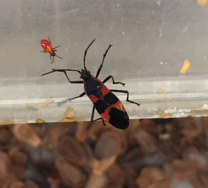
Holometabolist insects follow a lifecycle of egg to larvae to pupae to adult, while hemimetabolist insects develop from an egg to a nymph to adult. One example of hemimetabolism is in the Large Milkweed Bugs.


Schneider_LrgMilkweed_photo_galler_2

Large Milkweed Bugs (Oncopeltus fasciatus) exhibit incomplete metamorphosis. The nymph, or younger bug, resembles the fully grown adult only smaller in size.
Insects may pass through seasons in the egg stage; some grasshoppers have been known to pass through summer droughts in the eggshell, becoming dry and shriveled, and resuming development once moistened (Insects, 2015). Some may pass the winter in a diapause, a specific type of dormancy, in their egg and resume after the winter (Insects, 2015).

Schneider_CricketEgg_photo_galler_4

A zoomed in view of a cricket egg. Once the egg hatches a nymph will emerge that will grow and look similar to an adult cricket, only miniature. They go through incomplete metamorphosis.
After a holometabolist insect hatches, it enters the larval stage. Throughout this stage a gland in the larvae’s brain secretes neotenin, or juvenile hormone, which delays the metamorphosis from larva to pupa, the pupa developing when the amount of neotenin secreted is at a minimum (Insects, 2015). Similarly, neotenin also acts to prevent the metamorphosis from nymph to adult. As long as neotenin is present in the blood the molting cells lay down a larval cuticle, and in the final larval stage, neotenin is no longer produced, allowing the transformation from nymph into adult to begin (Insects, 2015). This allows for evolutionary success because many insects are able to inhabit more than one niche throughout their lives.

b. Throughout evolutionary time up to extant insects

The oldest known insect fossil, Rhyniognatha hirsti, dates back to the Devonian period. In many insect fossils, evidence exists of their feeding behaviors, such as feeding damage on vegetation and wood, giving clues to their habitats and food sources (Grimaldi and Engel, 2005). Insects have always been able to rapidly adapt to environments and resultantly can fit into many niches. Insect evolution is also closely tied to the evolution of plants, with up to 20% of extant insects relying on flowering plants, pollen, or nectar for food (Insect Evolution, 2007). Insects have not left a particularly strong fossil record, because with the exception of in amber, insects are mainly terrestrial and only preserved
in specific conditions on the edge of lakes (Insect Evolution, 2007)

IMG_5225

A 24 cm. Madagascar Hissing Cockroach (Gromphadorhina portentosa). Cockroaches are a great example of an insect that has changed relatively little since they first started appearing.

Remarkably, throughout time, insects have changed relatively little in that an entomologist today could likely identify many species buzzing or flitting around in the Jurassic period. Certainly it is millions of years of success that have prompted many to refer to our age as the age of the insects (Insect Evolution, 2007).
Extant Diversity

a. Abundance and Distribution
Due to the overwhelming success of hexapods in an evolutionary sense, extant or living hexapod abundance is astronomically high. Their high reproductive rates combined with their high variety make them present in extremely high numbers that are almost too large to comprehend. Some studies have shown that in one square yard of soil, up to 2,000 different insects can be found. This means that in just one acre there can be about 4 million different insects. Not only that, but hexapods have adapted to even the most extreme environments that range from deserts to the coldest mountains (Insect, 2015). One member of this extant group is the Australian Stick Insect, a bug that is extremely well camouflaged in its natural environment.

Schneider_WalkingStick_photo_galler_1

A female Australian Stick Insect (Extatosoma tiaratum) hanging from a small branch. The unique outer layer of the body serves to camouflage the insect.

Schneider_WalkingStickPlant_photo_galler_3
A zoomed out view of the Australian Stick Insects hanging from small tree branches.

Another extant member is the Praying Mantis, an insect that exhibits many of the defining traits of the subphylum hexapoda.

http://youtu.be/MC3inhf3CL4

A praying mantis (Mantodea burmeister) shows off his mouth parts and movement of his compound eyes.
The subphylum hexapoda can be broken down into various clades. First, there are the basic insects known as Class Insecta. This group makes up most of the winged insects that have been studied. According to Dr. Terry Erwin, an entomologist at the Smithsonian museum, this class may make up nearly 90% of all documented species on the planet (Erwin, 1982). Global populations of extant insects are estimated to range from 4 to 6 million different species (Novotney et al., 2002). Members of Class Insecta have also been found to be very adaptable, as shown by the fact that they are found in nearly every freshwater or terrestrial habitat. Some members have even found to be living in marine environments, though this habitat is primarily found to be home to crustaceans (Myers, 2001). There are also 3 groups of wingless insects or Class Entognatha: Collembola, Diplura, and Protura. Collembola, or springtails commonly, are the largest group and can be found essentially anywhere where soil or soil related habitats are found (Ponge, 1993). Populations of over 750 million individuals have even been found in certain grassland regions (Collembola, 2009). The groups Diplura and Protura are still abundant, but not nearly at the scale of Class Insecta. According to Dr. John Meyer, Diplura have around 800 species recognized and Protura have around 500 species recognized. Diplura are also common in grassy areas, but due to their small size they can sometimes be overlooked, meaning species estimates might be lower than reality (Diplura, 2009). Protura are less commonly found and can usually be associated with soil habitats that are damp or moldy (Protura, 2009).
b. Economic Importance
Hexapod insects can have a profound impact when it comes to the human economy. One of their biggest impacts come from natural resources. Insects can be used as a food source or can help produce other products. These products include can includes dyes and waxes, but the two primary products used by humans are honey from honey bees and silk from domesticated silkworms. Honey bees are also important due to their previously mentioned ability to pollinate plants, including human crops. Certain hexapods also have the ability to damage human products, such as wood homes that can be destroyed by insects like termites or furs and skins that can be broken down by insects like tineid moths. Hexapods also have the ability to help crops by assisting in pollination, or competing with humans by devouring said crops due to the herbivorous nature of most insects. Hexapods have a very important role in the medical community, due to the fact that many insects can be vectors, or carriers, for harmful microorganisms that causes diseases. Some of these diseases include malaria, yellow fever, sleeping sickness, and typhoid (Insect, 2015). This makes hexapods and other insects a large focus in biological medicine in order to help prevent such tragic illnesses. On that note, certain insects may also be used to assist in genetics research as well. The common fruit fly (Drosophila melanogaster) is one of these organisms. Due to its short generation time and high rate or reproduction, the fruit fly is considered to be a model organism that is used to study links between genes, evolution of behavior, and development in eukaryotic organisms (Pierce, 2006). Finally, insects can be a food source for human populations. Due to their high protein amounts, some believe that insects may have a high potential to be used in human diets, although diets that include insects are seen as abnormal in some higher developed countries (Gullan, 2005).

Conclusion

As our blog shows, hexapods are more than just the creepy crawly critters you squish under your feet. They have a rich evolutionary history dating back as far as 412 million years ago. Being one of the first insect groups to inhabit terrestrial habitats, hexapods served as a very important role in selecting land based plant life. Hexapods have many interesting characteristics including, the hexapod bauplan, wings, metamorphosis, and highly developed sensory systems.
If you still are not convinced that hexapods are a fascinating taxa of animals, don’t forget they are one of the largest and most diverse groups of animals on the planet. There are over 750,000 species of these guys! Hexapods ensure the success of many ecosystems, and we benefit from a lot of the ecological work they do. If you have ever stopped to admire a beautiful flower, chances are you have a hexapod to thank. Don’t worry if it is still hard for you to see insects as friendly little helpers, but we hope this blog gives you the insight to appreciate how amazing and important they are for our planet.

Works Cited
Clarke, D.; Whitney, H.; Sutton, G.; Robert, D. (2013). “Detection and Learning of Floral Electric Fields by Bumblebees”. Science 340 (6128): 66–9.

Erwin, Terry L. (1982). “Tropical forests: their richness in Coleoptera and other arthropod species”. Coleopt. Bull. 36: 74–75

Grimaldi, David; Engel, Michael S. (2005). Evolution of the Insects. Cambridge University Press. ISBN 0-521-82149-5

Gullan, P.J.; Cranston, P.S. (2005). The Insects: An Outline of Entomology (3 ed.). Oxford: Blackwell Publishing.

Insect. (2015). In Encyclopædia Britannica.

“Insect Evolution”. Virtual Fossil Museum. 2007.

Meyer, John R. (2009). “Collembola”. North Carolina State University.

Meyer, John R. (2009). “Diplura”. North Carolina State University.

Meyer, John R. (2009). “Protura”. North Carolina State University.

Myers, P. 2001. “Insecta” (On-line), Animal Diversity Web. Accessed April 20, 2015 at http://animaldiversity.org/accounts/Insecta/

Novotny, V., Basset, Y., Miller, S. E., Weiblen, G. D., Bremer, B., Cizek, L., & Drozd, P. (January 01, 2002). Low host specificity of herbivorous insects in a tropical forest. Nature, 416, 6883, 841-4.

Pierce, B.A. (2006). Genetics: A Conceptual Approach (2nd ed.). New York: W.H. Freeman and Company. p. 87.

Ponge, Jean-François (1993). “Biocenoses of Collembola in atlantic temperate grass-woodland ecosystems” (PDF). Pedobiologia 37 (4): 223–244.

Speight, Martin R.; Hunter, Mark D.; Watt, Allan D. (1999). Ecology of insects: concepts and applications (4(Illustrated) ed.). Wiley-Blackwell. p. 350.

Echinoderms Part Two

Echinoderm Environment:

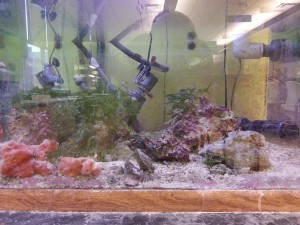
Echinoderms are the largest phylum with no freshwater or terrestrial forms. Echinoderm environments must be marine, as in saltwater, for the echinoderm to survive. Within marine environments, the conditions echinoderms live in can vary greatly. Environments range in water temperature, water depth, water movement and the different organisms surrounding the echinoderms. Water temperatures can range from arctic temperatures to tropical temperatures. Water depth and movement somewhat tie in together in the idea that the more shallow the water, the more likely the water will be moving. Deeper water is sometimes less affected by rain and storms and therefore does not affect the echinoderms as much. Water movement could impact the echinoderms by moving them or destroying their habitats. Depending on the species the water depth and movement will vary. Echinoderms are generally found in shallow water near shores or in reef environments but can also live in great depths of water. Almost all echinoderms are benthic, meaning they live on the seafloor, but some sea lilies can swim for some distance while some sea cucumbers float throughout the water (Xiaofeng, Hagdorn, & Chuanshang, 2006). Below is a photo of a tank for marine animals. This tank contains brittle stars, sea cucumbers, and sea urchins along with non-echinoderm animals. This marine tank is kept in conditions to reflect the echinoderm’s natural habitat. The tank is salt water and is kept at 78°F. In the tank are rocks, plants and other things to help give shelter and protection to the animals.

picture 1

This picture is of a tank in a classroom at Ohio State University. In this tank there is a species of brittle stars, sea urchins and sea cucumbers. There are also other animals such as a shrimp and some small fish. Not to mention the plants and sponges present.

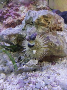
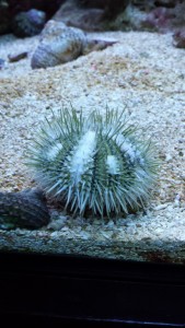
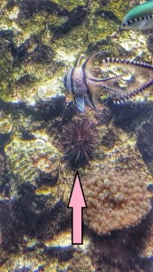
Brittle stars (Ophio coma) lead a cryptic lifestyle. They avoid predation by spending much of their day hiding within holes in coral and rocks found in their environment (Zubi, 2015). In the picture shown below, a brittle star is almost completely concealed under a sponge. Likewise, sea urchins’ main defense from predators is creating a burrow in the ground to leave less of their body exposed to predation. When sea urchins are found in environments not conducive to burrowing, like the marine tank shown above, they have been know to cover themselves with objects found in the environment for protection (Carefoot, 2010). The sea urchin shown below is exhibiting this trait, covering itself with shells from other organisms.

picture 2picture 3

The photo on the left is of a brittle star hiding out under a sponge in the tank at Ohio State. The photo on the right is of a sea urchin in the same tank carrying around a shell of another organism.


Lifecycle:

Though many Echinoderm species undergo sexual reproduction, a few species reproduce asexually. The bulk of echinoderm species are diecious, meaning that there are male and female individuals. The type of reproduction that an echinoderm goes through depends on the species and the environment (Mulcrone, 2005).

Sexual Reproduction:

Sexual reproduction refers to the combination of genetic material from two separate individuals, of different sexes, resulting in a new organism. The males provide the sperm and the females provide the eggs for reproduction. The sperm and eggs are propagated into the water column, and fertilization generally happens in the open water. Certain species of sea stars, brittle stars, and sea cucumbers go through internal fertilization. This is a recent observation and little information is known about this phenomenon. The release of the eggs and sperm is synchronized in two different ways. Some species have a breeding season where individuals congregate to increase the chances of fertilization. Some species’ breeding season may be affected by the lunar cycles. Commonly, echinoderms will leave their eggs and provide no parental care to the eggs (Young & Eckelbarger, 1994). Some species such as the Slender Sea Star (Leptasterias tenera) brood their eggs. This means that they carry their eggs with them until they are ready to hatch. This species of starfish carries the eggs in their stomach but other echinoderms carry the eggs in places such as in sacks under their arms, their coelom (main body cavity containing the organs) or in special egg cavities (Hendler & Franz, 1982).


Development:

When sexual reproduction occurs, the resulting eggs go through either direct or indirect development. When indirect development occurs, the fertilized eggs of echinoderms will develop into larvae known as planktonic larvae. In most cases, this stage occurs when the fertilized egg consists of a lower yolk volume.  The resulting larvae ends up assimilating into the surrounding plankton community. They will start feeding on numerous smaller organisms in an effort to meet the energy requirements needed to perform metamorphosis, and achieve their juvenile stage of their adult form. After a period ranging from one to three weeks, these bilateral symmetric larvae begin their metamorphosis, during which they will develop five water vascular canals that will eventually give them their characteristic pentaradial symmetry.

In contrast, during direct development, fertilized eggs will contain a larger yolk volume. Due to this, they are able to be sustain their life cycle development solely on their yolk, saving them from expending energy feeding, and also allowing them to bypass the larval stage of development. In this developmental strategy, often times the female parent will be involved in rearing the juveniles.  In these instances, the female will hold the young juveniles near its mouth, on the underside of its body, or even in specially developed pouches that are found on the upper side of the body. This allows the female to provide predation protection to its young until they fully developed. (Pawson, 2014)


Asexual Reproduction:

Echinoderms are capable of performing asexual reproduction, and the most common way they achieve this is through division of body parts or body sections, which is called fragmentation.  What happens in this reproductive strategy is that an echinoderm may deliberately or accidently detach a limb or body section. This may happen due to possible predation, an unforeseen accident, or in some cases with species of starfish, it separates itself by pulling opposing limbs in opposite directions until the individual splits. (Pawson/Encyclopedia Britannica, 2014)


The reason this works without permanently harming the individual is because of a unique process in which they are able to seal areas of exposed tissue where a section has been detached, and then immediately it begins regenerating new tissue to replace that section. Not all classes accomplish this strategy in the same manner, as multiple solutions seems to have arisen within each class. In species of starfish and brittle stars, this process requires that part of the central disk be included with each separate segment in order to regenerate into a complete individual. In species of sea cucumber, not only do they seal off the “wound” from where they split in half, the resulting two regenerating parts have to go through a tissue reorganization in order to successfully complete the process. (Speer and Waggoner 1999) In sea lilly and other starfish species, they can simply detach a limb or body section regardless of what part is present and either regenerate the missing limb, or in the limbs case, regenerate an entire new body.  While impressive, this asexual reproductive method has shown itself to be pesky for habitat management teams, as complete removal of sea stars and sea lilies can prove to be very difficult.  (Pawson/Encyclopedia Britannica, 2014)  


Echinoderms throughout evolutionary time:


The first true echinoderms appear in the fossil record in the Cambrian period, which began about 560 million years ago. During the pre-Cambrian period, echinoderm ancestors are not believed to have had radial symmetry (Zamora, 2012). Echinoderm ancestors in the pre-Cambrian are also thought to have been soft-bodied with unmineralized plates, which contrasts with all true echinoderms, starting in the Cambrian, who have hard mineralized skeletons which provide structure and protection (Speer, 1999). The earliest echinoderms are thought to have been deposit feeders, which intake and extract nutrients from the sediment on the floor of the ocean. This varies from the current method of feeding found in echinoderms today, suspension feeding, in which echinoderms eat particles found suspending in their environment (Steele). The first echinoderms in the Cambrian period had both radiate and asymmetric forms and a similar water vascular system to those found today, including one coelom (Zamora, 2012).


During the mid-Cambrian, the species richness, or the number of species, increased by fifteen percent. In the Ordovician period, starting around 510 million years ago, the earliest echinozoans, starfish, and brittle stars appear (Baumiller, 2015). After the Great Ordovician Biodiversification Event (GOBE), suspension-feeding organisms, like the echinoderms dominated ocean floor habitats. Three echinoderm classes from the Cambrian went extinct during this period, six classes survived from the Cambrian, and eleven new classes appeared after the GOBE. No new classes of echinoderms have emerged since the GOBE (Baumiller, 2015).


Since the Ordovian period, the number of classes has steadily declined. Though the classes of sea lilies and blastoids dominated the echinoderm phylum through most of the Paleozoic period, by the end of the Permian period, which ended 250 million years ago, all of the blastoids and almost all of the sea lilies were extinct. From the Mesozoic period, which directly followed the Permian period, to today, there have been only five classes of echinoderms, the Sea Lilies (Crinoidea), Starfish (Asteroidea), Brittle stars (Ophiuroidea), Sea Urchin (Echinoidea), and Sea Cucumbers (Holothuroidea) (Baumiller, 2015).


Diversity:

With 7,000 living species, echinoderms are very diverse (Mulcrone, 2005). Echinoderms vary in physiology and morphology such as shape, size (width and length), color and locomotion. Amongst the five main classes of echinoderms, probably the most obvious difference is the shape of the body. For example sea stars have a star looking shape while sea urchins are spherical, sea biscuits and sand dollars are round, and sea cucumbers are longer and more caterpillar-like. Below is a photo, from left to right, of a brittle star skeleton, sea cucumber and sea urchins remains, followed by live specimens of sea cucumbers, sea urchins, sea biscuits, and sand dollars. The diversity in the body shape can clearly be seen.

sea cucumber


urchin1 urchin2 urchin3 urchin4

In the photo above you can not only see the diversity in body shape but also the color and size. The brittle star in the photo is an Orphiarachnella ramsayi and was found on a reef in Sydney Australia. These brittle stars can range in colors from black and white to having some pink, red and green as well (O’Hara, 2015). Compared to the sea cucumber that is bright orange, the color obviously varies. The size between these three specimens can range from 1-10 inches. Diversity in echinoderms is not only between the five classes but between the species in each class. For example sea stars vary in size, color and number of appendages. Below is a photo of three very different sea stars.


starfish
This photo shows the variance in size, color, and number of limbs that can be on just sea stars alone. The starfish on the left was about seven or eight inches across while the starfish on the right was less than an inch.  The color variation is also shown.

blue starfish

Shown above is a blue sea star, Linckia laevigata.


As seen in the photo the sea star on the left is larger and has only five appendages while the sea star on the right is much smaller and has six appendages. The sunflower starfish (Pycnopodia helianthoides) can have between 16 and 24 arms and can be up to a meter in diameter (Sustainability Species Identification). This just shows the diversity of appendages in sea stars. The sea cucumber and sand dollar have no appendages at all. One other notable difference that has occurred between echinoderms is their locomotion, or how they move. For example sea stars move by using their tube feet to attach to the ground and sort of walk. Brittle stars on the other hand use their appendages to push themselves from place to place. Finally, sea cucumbers can pull themselves along somewhat like a snail and even some species can swim (Mah, 2012). Sea urchins, primarily move using their tube feet, however, they can also use their spines for fast movement, as shown in the video below. The next two videos show a brittle star, a chocolate chip sea star, and a sea urchin moving.


This video shows how a brittle star uses its limbs, as opposed to tube feet, to move around the tank.


This is a timelapse of a starfish moving around on top of an oyster.

https://www.youtube.com/watch?v=s7qagaFoHyk&feature=youtu.be

This video shows a long-spined sea urchin, Diadema setosum, moving using its spines. Sea urchins primary mode of locomotion is their tube feet, but they occasionally use their spines for movement.

This is just a short overview of the diversity of echinoderms that has developed over millions of years.


Human impact:


Human beings as we continue to grow in numbers have a lasting impact on many different animal species, however, it is becoming more evident of our impact on the oceans and the animal species that call the oceans home. This section will specifically cover the effects humans have on echinoderms but many of these effects have a cascading effect on many different animals.


The fishing industry has a large impact on the survival of echinoderm species. The fishing industry removes substantial amounts of the echinoderms’ diet from the oceans including, clams, mussels, and oysters. This reduces the food available to the echinoderms. The pet trade also has several direct effects on many species of echinoderms. A big part of the pet trade industry involves collecting corals and live rock for sale in many countries all over the world. The coral and live rock make up the habitats of numerous echinoderms and removing them effectively leaves the echinoderms unable to protect themselves. Also, humans pull all echinoderms (starfish, sea urchins, sea cucumbers, sea lilies and brittle stars) out of their natural habitat to be directly sold in the pet trade. Both of these problems affect the number of echinoderms in the wild. Taking rock from reefs removes a lot of habitat that these animals rely on, and removing them from areas completely diminishes the number of breeding animals.


Humans play a large role in the habitat destruction affecting many echinoderm species. Human have a large carbon footprint, and as we are releasing large amounts of carbon dioxide into the atmosphere, the pH of the oceans is slowly rising. This slight increase in pH is not detrimental to adults in all species currently, but it has a significant impact on the more sensitive species of echinoderms. Also, these environmental changes do affect all echinoderms during their developmental stage when they are the most susceptible to environmental changes. This effect can be severe; a change of just 0.2 units of pH leads to a 100% mortality rate in 8 day old common brittle stars (Ophiothrix fragilis) (Dupoint, 2009).


Conclusion:

Echinoderms are found all over the world and play a major role in the animal kingdom and the environment. They help to keep algae growth down, feed other animals, and feed people in certain countries. People need to understand the importance of echinoderms. If too many are captured or we keep destroying their habitats, it could affect more than just the echinoderm’s future. As a keystone species in many ecosystems, the loss or elimination of species of Echinoderms could drastically and permanently affect numerous marine habitats around the world, as well . They are a diverse family, with 7,000 currently known species of echinoderms ranging from sea stars to sea cucumbers, and with the scientific community making new discoveries using ever developing technology, we could see many more species discovered in our lifetime.


Work Cited:


Baumiller, T. (2015). Fossil record of Echinoderms. Retrieved from https://scripps.ucsd.edu/centers/echinotol/about-echinoderms/fossil-record-of-echinoderms/


Carefoot, T. (2010). Learn About Sea Urchins: Predator and Defenses. Retrieved from http://www.asnailsodyssey.com/LEARNABOUT/URCHIN/urchDefe.php


Dupoint, S., & Thorndyke, M. (2009). Impact of CO2 -driven ocean acidification on invertebrates early life-history – What we know, what we need to know and what we can do.Biogeosciences Discuss, 6, 3109-3131. accessed 29 March, 2015.


Echinodermata. (n.d.). Retrieved from http://www.bio200.buffalo.edu/labs/echinoderms.html


Echinoderms. (n.d.). Retrieved from http://www.oceaninn.com/wildlife/echinoderms.htm


Hendler, G and Franz, D. (June, 1982). Washington, DC: Smithsonian Institution and New York, NY: Brooklyn College.


Mah, C. (2012, Sept. 18). Deep-Sea Swimming Sea Cucumbers and the “most bizarre holothurian species in existence”! Retrieved from http://echinoblog.blogspot.com/2012/09/deep-sea-swimming-sea-cucumbers-and.html


Mulcrone, R. (2005). Echinodermata (sea stars, sea urchins, sea cucumbers, and relatives). Retrieved from http://animaldiversity.org/accounts/Echinodermata/


Pawson, D. (2014, July 24). Echinoderm. Retrieved from http://www.britannica.com/EBchecked/topic/177910/echinoderm


O’Hara, T. (2011). Brittle Star. Retrieved from http://portphillipmarinelife.net.au/species/6607


Speer, B., & Waggoner, B. (1999, September 15). Echinodermata: Fossil Record. Retrieved from http://www.ucmp.berkeley.edu/echinodermata/echinofr.html


Speer, B., & Waggoner, B. (1999, September 15). Echinodermata: Life History and Ecology. Retrieved from http://www.ucmp.berkeley.edu/echinodermata/echinolh.html


Steele, M. (n.d.). Ecology of Soft-Sediments. Retrieved from http://www.csun.edu/~msteele/classes/marine_ecology/lectures/15_soft-sediment ecology.pdf


Sustainability Species Identification. (n.d.) Sunflower sea star. Retrieved from http://www.nmfs.noaa.gov/speciesid/fish_page/fish6a.html


Xiaofeng, W., Hagdorn, H. and Chuanshang, W. (2006). Pseudoplanktonic lifestyle of the Triassic crinoid Traumatocrinus from Southwest China. Retrieved from http://onlinelibrary.wiley.com/doi/10.1080/00241160600715321/abstract;jsessionid=9342400ED743C5DFC4E653363D1CDAF0.f02t02


Young, Craig M.; Eckelbarger, Kevin J. (1994). Reproduction, Larval Biology, and Recruitment of the Deep-Sea Benthos. Columbia University Press.


Zamora, S., Rahman, I., & Smith, A. (2012). Plated Cambrian Bilaterians Reveal the Earliest Stages of Echinoderm Evolution. PLoS ONE, 7(6).


Zubi, T. (2015). Invertebrates: Echinoderms. Retrieved from http://www.starfish.ch/reef/echinoderms.html


Molluscs Part 2

Where & How Do They Live?

Though under the same Phylum, each of the major classes discussed in this blog live extremely different lifestyles. Their diet, anatomy, locomotion, reproduction, habitat, and life history all differ remarkably, even though they share many synapomorphies. Gastropods, like our garden snail, play it safe and slow, primarily filter feeding and seeking protection in their shell or in other safe environments. Bivalves, such as the mussels, diverged from their mobile ancestors in order to live a sessile life, but still manage to feed and reproduce with extreme success. Cephalopods, like the squid, are the hunters of group, as they have derived tentacles along with sharp muscular chitin beaks in order to catch and process food.

Unique Anatomy

Around the Devonian period, bivalves with siphons appeared. According to Nordsieck (2011), the addition of siphons along with the bipartite shell development, afforded bivalves to exhibit extraordinary protection allowing them to only need to extend their siphon in order to breathe, to feed, and to reproduce, without having to expose the rest of its body. During the Mesozoic period, burrowing bivalves with siphons underwent some species differentiation that eventually proliferated into other time periods. For example, Nordsieck (2011) point out that swimming scallops appeared during the Triassic, reef building Rudist bivalves dominated during the Cretaceous displacing coral and freshwater bivalves appeared in the Devonian.

In Gastropods, the shell is very different from other mollusc shells as it is coiled to form its characteristic spiral. Garden snails for example evolved to have developed a dorsal sack, known as the visceral hump, to contain most of the internal organs. This part remains under the mantle and is always within the shell for maximum protection. During embryonic development “torsion” occurs, as the mantle and the visceral hump turn around and coil into the spiral saving space, meaning that gastropod shells are coiled asymmetrically to one side depending on this torsion . Because of the twisting of the digestive tract, the anus in Gastropods is located above their head (Nordsieck, 2011).

Cephalopods are the most derived mollusc group. They reside in the subphylum Conchifera, containing mainly molluscs that have retained their shells. Which is weird seeing as cephalopods have lost their shells completely. They demonstrate a body plan similar to that of slugs and other unshelled gastropods: a reduction of the shell, at the cost of protection but improving movability. However, Nautilus, an extant species of cephalopods, still bears an external shell. Nordsieck (2011) mentions that squids also have a highly developed nervous system,and contain chromatophores, which can change the color of their skin cells.The unique adaptation gives cephalopods ability to visually communicate with one another, as well to camouflage themselves.

Marine Vs. Freshwater Vs. Terrestrial

Bivalves are aquatic and are mainly found in marine environments, however, multiple bivalve groups have adapted to freshwater environments several times, resulting in several non-closely related freshwater mussel groups according to Nordsieck (2011). Because these freshwater environments sometimes have no water flow, bivalve larvae and are often adapted to attach to mobile inhabitants, as the case with the fingernail clam, in order to disperse itself throughout the environment.

According to Nordsieck (2011) gastropods are unique in that they are the only of our three classes to live in marine, fresh-water, and terrestrial environments! How cool!. Their shell plays a large part in their adaptation to terrestrial environments, as it provides protection against desiccation by trapping water and by giving mobile protection against solar radiation, as seen in our model species the Brown Garden Snail.

Cephalopods are restrained to only marine environments. This is because of their large, developed nerve system that is strongly correlated with the salty environment. According to Norman (2013), “It is probable that they never developed a sodium pump that would help them cope with osmotic change in freshwater.” Even if they did, they would be much more subject to predation than in the open ocean, and would be severely disadvantaged.

Locomotion

Though largely sessile, Bivalves still have the ability to move short distances. The most common method of moving based on studies by Nordsieck (2011) is reaching their muscular foot from out of their shell, anchoring it to a nearby substrate, and then contracting the foot pulling the Bivalve towards the tethered end. Studies done by the Cambridge University Museum of Zoology (2011) show that the foot is also used in a similar way to burrow into sediment. This allows it to stay anchored in one place while filtering, so that it does not get dragged away by water currents. Though this is a common form of locomotion, the Blue Mussel is actually quite different. According to the Living World of Molluscs (2011) these mussels throw out a thread to attach to something in the environment, then slowly shrink the thread, drawing the Mussel to the attachment as if it were a “grappling hook”.

Holthuis (1995) makes the point that gastropods are actually very mobile in comparison to bivalves, as they crawl along on their large, flat, muscular foot. Their foot contains glands that secrete a mucus, which facilitates this movement, leaving their trademark slimy trail (Dekle & Fasulo, 2015).

Cephalopods are incredibly mobile, and are by far the most agile taxon in the phylum. As stated by Howard (2003), they use jet propulsion in order to move. This involves pumping water through their bodies and out of a tube known as the siphon. The siphon is a muscular organ it uses to steer itself by directing the water flow in order to control the direction of propulsion. Some bivalves have adapted a similar method of jet propulsion, allowing them to launch themselves through the water!

Feeding

Bivalves are completely dependent on filter feeding, meaning that they feed by using their muscle to pump water into their body through their siphon, and then sifting out particles for nutrition. Nordsieck (2011) states that edible particles are transported to their digestive tract, and inedible particles are excreted through the exhalant siphon. This ability to improve the quality of water is an incredibly crucial ability that is essential to maintaining water quality around the world. However, it is because of this feeding method that bivalves are so afflicted by sea water pollution, and will accumulate harmful substances in polluted areas.

Gastropods are primarily herbivores, relying on their shell as a protection in order to slowly explore open environments to reach rocks and other hard substrates which may contain food. Nordsieck (2011) says that in aquatic environments, gastropods primarily feed on alga, but in terrestrial environments, snails such as the Brown Garden Snail primarily consume herbs and vegetables. This alga consumption makes them important organism in any aquatic environment, as they play a large part in keeping alga levels from rising too high, which could be harmful towards the ecosystem. They also assist in breaking down dead tissues by using their rasping tongue to scrape away at the substrate, assisting in the process of decomposition.

Cephalopods have a highly derived foot used primarily for hunting, which works well with their carnivorous diet. The foot has been modified into arm-like tentacles, which contain suckers that are used while hunting for grabbing prey (figure 7), and then restraining it as the beak crushes the prey while the radula tears and ingests the tissue. As stated by Howard (2003), cephalopods are also the least dependent on a solid substrate to move, and so are able to catch prey unlike the herbivorous and filter-feeding bivalves and gastropods. Besides fish, squids’ prey are other molluscs as well as crustaceans.4 - Ondrejech_Andy_Cephalopod_Foot_Photo

Figure 7 – An Up-close picture of the suckers on an octopus tentacle. This is used to restrain prey while hunting and feeding.
* Sucker is approximately 1-2 cm wide

Reproduction

A detailed account by Nordsieck (2011) describes the Blue Mussel life cycle. They have separate sexes, and use the water they are so dependent on to reproduce by releasing 5 – 12 million eggs into the water per female! The males also release sperm, in hopes the two will meet in a chance fertilization. A larval stage will develop from this, and may be transported several hundred kilometres by sea currents. Most (about 99.9%) of mussel larvae will serve as food to other sea creatures, but some will survive and attach themselves to algae. Until they have reached the size of 5 cm, they will move about until they finally settle to a suitable piece of ground. Preferably, this spot is by other mussels, forming a mussel bed. This is preferable because the chance of fertilization is much higher when around more mussels!

Though most bivalves have separate sexes, there are some hermaphroditic groups. This occurs in oysters for example. It occurs when the number of male oysters is too high in comparison to the female population, resulting in some male oysters altering their sex to become female! There are some bivalves in which fertilization occurs in the female’s mantle cavity, and then develop into a parasitic larval stage which must infect a fish in order proceed to become young mussels.

Gastropods are also very diverse when it comes to reproductive strategies! Though it varies widely, there are some correlations between method and the environment. Nordsieck (2011) states that Aquatic snails often use the water to transfer sperm cells. Terrestrial snails have sperm packets known as spermatophores that are not dependent on water and resistant to desiccation. In the case of the Brown Garden Snail, individuals are hermaphroditic, meaning that they have the reproductive organs of both sexes. Dekle & Fasulo (2015) state that, thanks to this trait, snails are can fertilize themselves, even though cross fertilization is common, meaning that they fertilize each other at the same time! Ovoviviparity is also found in both terrestrial and aquatic snails, meaning that the eggs hatch inside the mother’s body and are born alive!

Cephalopods are unique in their phylum because they are only male and female! Male squids have a special arm called a hectocotylus, which is slightly shorter than the rest. According to Nordsieck (2011) this arm is used to internally fertilize females’ by transferring a sperm packet to the female’s mantle cavity. This fertilizes the female’s eggs, which are often in the thousands. These are then hidden where they develop into their larval stage, but unfortunately, most are eaten before they can grow large enough to avoid being such an easy meal! In all taxon, larvae are a very important part of the food chain. Without the massive amount of larvae that become food for other marine species, many species may not persist!

Extinct Taxon

Each class consists of many groups of Molluscs, but many of these groups have gone extinct. Harvard Research Site (2010) states that among these were the Rudists, which were ring-shaped bivalvia important to the formation of reefs, and Ammonites, which were heavily shelled, predatory cephalopods that fed on plankton in open ocean water.

Though there once ten classes of Molluscs, two of these classes, Rostroconchia and Helcionelloida, are both extinct. According to Pojeta (1972), though the Rostroconchia were a sort of filter feeding, bivalve-like creature, they did not have many similarities with the bivalves that we know and love. Actually, they are considered to be more related to Tusk Shells, which is a phylum that we did not discuss in this blog. Despite having over a thousand species they are believed to have gone extinct in the Permian Extinction.
Helcionelloida resembled snails, as they had a large muscular foot, as well as cone-like shell with large ridges on it. Though in it’s own class, the Czech Geological Society (2011) have reason to believe that Helcionelloida may possibly be the ancestors of Cephalopoda, Bivalvia, and Gastropoda.

Survey of Extant Taxa

Distribution and Abundance

As molluscs are so diverse, it makes sense that they are found in nearly every ecosystem. Mostly molluscs are marine, inhabiting corals, beaches, shores and other habitats. They can also be found in the deep ocean, with the Giant Squid being the most recognizable example. In addition, the diverse gastropod class has provided terrestrial mollusc species, which are able to inhabit most terrestrial ecosystems (Bunje, 2003).

Gastropods (such as the Garden Snail picture below) range second, only behind insects when it comes to the number of named species. They make up over 80% of all living molluscs and are one of the most highly diversified classes in the phylum according to Myers & Burch (2001). Today there are more than 62,000 living species of gastropods. They live in different habitats and display extreme diversity in their morphology, whether it’s dealing with their size, body, or shell. They are the only molluscs group to have invaded land habitats and thus they occupy the widest ecological niche of all molluscs. They are found in deep seas, freshwater habitats, salt lakes, mountaintops, deserts, rainforests and other habitats.Estimations for the extinct species range from 40,000 to 100,000 and some even believe it to be as many as 150,000 species. As Nordsieck (2011) states, gastropods have rich fossil record which goes back to the late Cambrian period, that is nearly 600 million years ago. These fossils also show both extinctions and diversification of new groups.

2015-02-09 15.35.08

Figure 8- Brown Garden Snail (Cornu aspersum)
*Approximately 1 ¾ in long

The Brown Garden Snail (Cornu aspersum)is a member of Helicidae family. According to Nordsieck (2011), “It is between 25 and 40 millimetres wide and between 25 and 35 millimetres high”. It has originated from western Europe, Britain and along the borders of the Mediterranean, but today it is spread worldwide. According to Dekle and Fasulo (2014) it has been introduced to places like North America, South America, Mexico, Australia, New Zealand and even parts of Africa.

The class Bivalvia is the second largest class, according to Vaughan, et al. (2014), among molluscs with about 10,000 species, and around one-fifth of those species believed to be freshwater lovers. Bivalves can be found at most depths in the ocean. In shallow waters, bivalves are found on sandy and rocky shores, and can often wash ashore. Bivalves are also found in the deep ocean, at abyssal depths. In these parts of the ocean, bivalves have been known to borrow and surface dwell. They are also found in all latitudes and depths in between: in soft shales, mud banks and coral reefs (Morton, 2013). Bivalves are first found in the fossil record in the Early Cambrian (about 542 to 509 million years ago) and diversification of bivalves weren’t found in the fossil record until the Early Ordovician (around 485-470 million years ago).

The Blue Mussel (Mytilus edulis) species are distributed in the Arctic, North and Mid-Atlantic, and North Pacific Oceans, although the blue mussel is native to the eastern coast of the United States. The blue mussel is a very abundant species in the North and Mid-Atlantic Oceans, and is abundant enough to be known as the “common mussel” (Newell, 1989).

In comparison to our previous two classes, the cephalopod class is a significantly smaller in size, only containing about 700 species. However, despite the small number of species, cephalopods vary greatly in size: they can get as big as 60 feet, as in the case of the Giant Squid, or as small as half an inch, like the pygmy squid (Seible, 2015). Cephalopods are found in every region of every ocean. They can also be found in all depths; some can be found in shallow to mid-level waters, where some others are bottom-dwellers (Snyderman, n.d.). There have been a recorded 15,000 species of cephalopod species, indicating the high diversity that they had experienced. In fact, cephalopods used to be so diverse that three entire clades that have gone extinct. The cephalopods first recorded in the fossil record originate from the Late Cambrian period, about 497-485 million years ago (Vendetti, 2006).

European Squids (Loligo vulgaris) are a cephalopod commonly found in deep waters in the southern North Sea, but voyage to coastal waters to spawn. They are also found in scattered areas from the North Atlantic/Norwegian Sea to the Mediterranean Sea and the coastal waters of northwest Africa (Telnes, 2014). The depth of the squid usually varies along with the temperature. The warmer the temperature is, the shallower they swim- usually at only a couple hundred meters. However, during colder temperatures, these squids usually can be found at depths as deep as 1000 meters. The European Squid is also an abundant species, being very common in the eastern Atlantic Ocean (Gibson, 2009).

Marine and Freshwater

As filter feeders, bivalves have become an important animal within their ecosystems. Originating from a marine, saltwater ancestor, bivalves have evolved to inhabit both marine and freshwater environments. Within the subclass Palaeoheterodonta has a freshwater-only order called Unionoida, and under the order of Veneroida (subclass Heterodonta), contains a three seperate freshwater families (Kohl n.d.). Based on the lineages, we can determine that the adaptation of living in freshwater water has occurred through parallel evolution in mussels (Morton, 2013).

For the class of molluscs, freshwater and terrestrial species often experience a higher rate of extinction than the marine species. In fact, around 99% of mollusc extinctions are non-marine species. There are many theories for this:human activities disrupting their habitat and the introduction of invasive species. Human habitat destruction could be a result of forest-clearing, pollution, construction of dams, or urban and agricultural development. Most molluscs, especially freshwater mussels are very sensitive to changes in water quality. In addition, invasive species can lead to alteration of the habitat, a competitive disadvantage to the invasive species, and predation and diseases from the new species (Parent, 2008). This can cause problems to native species and can potentially lead to extinction.

Conclusion

As we have shown, molluscs are an extremely diverse,important, and astounding species. Though it is difficult to pinpoint their exact evolutionary history, it is nothing less than incredible that the few synapomorphies such as the foot, radula, and shell could create such a diverse array of organisms that the world we know today could not exist without. According to Nordsieck (2011), a mussel can filter approximately two to three liters of water in a day (figure 9), enough to filter entire seas in just a few days! Without these organisms, what would keep the water that we and many other organism need clean to survive?

Molluscs have many direct benefits to humans as well. Squid ink is an important to cosmetics (Nordsieck, 2011), Cone Snail venom is being studied for it’s properties as a neurotoxin, and many molluscs can be exploited as a source of food (Harvard, 2010). Even the physical properties of molluscs have great value, as pearls produced from bivalves are important to jewelry, the shells of something strange molluscs are coveted and hunted, and in some ancient societies, the shells were even used as currency (Harvard, 2010). So whether it is gastropods decomposing tissues, squid larvae fueling fish populations, or the cultural importance of mollusc containing dishes such as escargot, molluscs are ingrained into our life as they are throughout the entire physical world.

Preserved Bivalve
Figure 9 – The Unique Internal anatomy of a Mussel is a key part of what makes it such an effective filter feeder.

Molluscs are also proven to be good indicators of ecosystem health. They are very vulnerable to changes in the environment, and some of the most endangered of all are some of the most important; the freshwater mussels. Sedimentation can bury mussel beds, streams can be clogged with algae from agricultural nutrient runoff, and pollution can harm populations (Morrison 2010). There is hope. Improved water quality and stream bank revetment efforts have stabilized some mussel habitats. New developments in mussel propagation techniques provide hope for these endangered species. In many ways, the fates of men and mussels are linked.


                                            Work Cited

1. Myers, P., & Burch, J. (2001). ADW: Gastropoda: INFORMATION. Retrieved February 12, from http://animaldiversity.org/accounts/Gastropoda/
2. Nordsieck, R. (2011). The Living World of Molluscs. Retrieved February 12, from http://www.molluscs.at/index.htm.
3. Morton, B. (2013). bivalve | class of mollusks :: Ecology and habitats | Encyclopedia Britannica. Retrieved February 12, from http://www.britannica.com/EBchecked/topic/67293/bivalve/35737/Ecology-and-habitats#toc35738
4. University Museum of Zoology, Cambridge | Burrowing Bivalves. (2011). Retrieved February 12, from http://www.museum.zoo.cam.ac.uk/bivalve.molluscs/lifestyles.of.bivalve.molluscs/burrowing.bivalves/
5. Harvard Library Research Guides. (2010). Retrieved March 23, from http://guides.library.harvard.edu/Mollusks
6. Dekle & Fasulo (2015). Brown Garden Snail, Cornu aspersum (Muller, 1774) (Gastropoda: Helicidae)1. Retrieved March 29, 2015, from http://edis.ifas.ufl.edu/in396
7. Vaughan, Dionne, Hui, Nguyen, Search, & Zhang (2014).Bivalvia Family Index. Retrieved March 29, 2015, from http://shells.tricity.wsu.edu/ArcherdShellCollection/Bivalves.html
8. Morton (2013). Bivalve | class of mollusks. Retrieved March 29, 2015, from http://www.britannica.com/EBchecked/topic/67293/bivalve
9. Bunje (2001).The Bivalvia. Retrieved March 29, 2015, from http://www.ucmp.berkeley.edu/taxa/inverts/mollusca/bivalvia.php
10. Newell (1989). Species Profiles: Life Histories and Environmental Requirements of Coastal Fishes and Invertebrates (North and Mid-Atlantic). Retrieved March 29, 2015, from http://www.nwrc.usgs.gov/wdb/pub/species_profiles/82_11-102.pdf
11. Seibel (2015). Science Explorations. Retrieved March 29, 2015, from http://teacher.scholastic.com/activities/explorations/squid/libraryarticle.asp?ItemID=213&SubjectID=113&categoryID=5
12. Snyderman (n.d.). Many Arms, Many Feats. Retrieved March 29, 2015, from http://www.dtmag.com/Stories/Marine Life/02-06-whats_that.htm
13. Vandetti (2006).The Cephalopoda. Retrieved March 29, 2015, from http://www.ucmp.berkeley.edu/taxa/inverts/mollusca/cephalopoda.php
14. Telnes (2014). European Squid – Loligo vulgaris. Retrieved March 29, 2015, from http://www.seawater.no/fauna/mollusca/vulgaris.html#
15. Norman (2013). Are there any freshwater cephalopods? Retrieved March 29, 2015, from http://www.abc.net.au/science/articles/2013/01/16/3670198.htm
16. Parent (2008). The Global Decline of Mollusks. Retrieved March 29, 2015, from http://www.actionbioscience.org/biodiversity/parent.html
17. Pojeta (1972). Rostroconchia: A New Class of Bivalved Molluscs. Retrieved April 19th, 2015, from http://www.sciencemag.org/content/177/4045/264
18. Czech Geological Survey (2011). Helicionelloida. Retrieved on April 19th, 2015, from http://muzeum.geology.cz/d.pl?item=99&l=e
19. Morrison (2013). Freshwater Mollusk Conservation Society. Retrieved on April 19th, 2015, from http://molluskconservation.org/

Angiosperms Part Two


 Lifecycle & Ecology


https://www.youtube.com/watch?v=j6LCCxFeDq8&feature=youtube_gdata

The Angiosperm Life Cycle Video – This is a look at the growth, fertilization, and pollination events in flowering plants. The diversity of morphology in angiosperm structures helps to facilitate this life cycle. It is the reason that angiosperms have been able to be ubiquitous throughout the various biomes of our Earth! Created by Emily Thomas.

Angiosperms are considered to be one of the greatest examples of symbionts in nature, due to their many mutualistic relationships with pollinators, fungi, herbivores and others. They can be found in almost any environment, so long as there is sunlight, some form of water, and a way to spread their offspring. The general life cycle of angiosperms is explained in the video on this page, ‘Angiosperm Life Cycle.’ While this video focuses on a more general view, the four major events that make up flowering plant reproduction (pollen development, egg development, pollination and fertilization) will be focused on in more detail here.  Angiosperms produce two types of spores; microspores which lead to the generation of pollen and megaspores which form the structure that houses female gametophytes (Boundless, 2014).

Pollen develops inside the stamen. Inside the anther of an angiosperm lie the diploid microspores. These microspores undergo meiosis to become haploid microspores. Haploid microspores then undergo mitosis to develop into pollen grains. Pollen forms from the male gametophyte in flowering plants.  The gametophyte consists of two cell types: tube cells to aid in fertilization and generative cells which generate sperm cells (Taiz, 2006).

Egg development occurs inside the carpel. Inside of the ovaries are egg producing structures known as ovules. Inside of an ovule are diploid cells. Similar to the production of pollen, these diploid cells divide via meiosis to become haploid cells that are the megaspores. These spores then go through three rounds of mitosis forming seven cells. One of the cells has two nuclei, called the endosperm and another of the cells becomes the egg. The remaining five cells are not used in reproduction (Boundless, 2014).

Once both eggs and pollen development have taken place they are ready for pollination. Pollination can occur in many ways; two major forms are wind and water dispersal. Unique to angiosperms is the use of pollinators such as birds and bees.  The last step in flowering plant reproduction is fertilization. For fertilization to occur the tube cell of the male gametophyte creates a tube to the ovule (Taiz,  2006). The generative cell uses this tube to send sperm down to the ovule so fertilization can occur. One sperm will fuse with the egg forming a zygote the other fuses with the endosperm forming a triploid endosperm cell. This process, known as double fertilization, is unique to angiosperms (Derksen, 2013). The endosperm later develops into nutrient tissue while the zygote divides by mitosis, developing into an embryo which grows into a mature plant.

Angiosperms spend most of there life in the adult stage known as a sporophyte. When we see trees, grass, flowers, vegetables in a garden we are seeing sporophytes! Angiosperms are very important due to their abundance and impact on almost every habitat on earth. Due to their diverse morphology they can range from the small to massive, aquatic to mountainous, grass to trees and everything in between.

A fruiting angiosperm. Photo taken by Nick White.

A fruiting angiosperm. Photo by Nick White.


 Evolutionary History


The Amorphophallus titanum, or Corpse flower is one of the most bizarre and oldest ancestors of modern angiosperms. Photo taken by Nick White

The Amorphophallus titanum, or Corpse flower is one of the oldest and most bizarre ancestors of modern angiosperms. Photo by Nick White.

Not surprisingly, angiosperms are the most commonly found type of land plant. Angiosperms evolved in the Cretaceous era, around the same time as many groups of modern insects. Many of these insects acted as pollinators that drove the evolution of both angiosperms and the insects themselves. (Soltis 2005). Due to the availability of pollinators (insects occupy nearly every environment on the planet) it has allowed angiosperms to become the most numerous plant found on land. This relationship is considered one of the greatest examples of symbioses in nature due to their many mutualistic relationship with pollinators, fungi, herbivores and others. They can be found in almost any environment, so long as there is sunlight, some form of water, and a way to spread their offspring.With co-evolution, these two species have been able to occupy places that few other species previously could, changing the habitats of the entire planet.(Lerner 2008)

Unlike many land plants, angiosperms did not evolve from gymnosperms. It is unclear what type of plants gave rise to angiosperms. (Angiosperms 2014) Some scientists believe that a group of plants known as “seed ferns” ,or pteridosperms, may have been the progenitor of the angiosperms. These “seed ferns” were around for many millions of years before angiosperms and yet have similar traits like seed-bearing capsules and specialized organs that produced pollen. While we are still not exactly sure how ancient angiosperms may have come about, we have an idea of what these ancestors may have looked like. They were likely small with small flowers. The flowers were probably green and not at all like the flowers we are used to as their sepals and petals would not be separated or distinguishable (Angiosperms 2014). And while the exact way that angiosperms evolved to what we know today is still unclear, their impact on our world today is obvious.


 A Survey of Extant Diversity


Angiosperms are arguably the largest extant group of plants on the planet today.  At least 260,000 living species exist, which are classified into 453 families (Soltis 2005). The most popular

The photo above are barrel cacti (Echinocactus grusonii) displaying the numerous spines that are used for protection. Additionally, these cacti have modified leaves, which help retain water in dry environments; they are also known as succulents. Photo by Nick White.

lineage would be the eudicots, which includes most flowering plants.  Some other major lineages are the Monocotyledons, containing families like lilies, grasses, and orchids, and the Nymphaeaceae, which hold the water lilies and their relatives (Soltis 2005).  Angiosperms inhabit all seven continents, as well as the oceans.  They are able to occupy just about any environment on earth, for example, high mountaintops, deep oceans, freezing tundras, and of course, warm, wet rainforests.  Their abundance in these environments is immense.  They have an extremely large genome, which may explain their ability to exist in so many different morphological forms. Some examples of these forms include grasses, climbing vines, large trees, and small flowers.  By diversifying their physiology, angiosperms have been able to adapt to the variety of ecosystems which cover the earth. The cactus, for example, has modified leaves, called spines, which help to prevent it’s desiccation in dry, arid deserts (see photo below).  Some types of angiosperms can be quite special and complex in terms of their nutrient acquisition, for example, carnivorous flowers, or poisonous vines.

Angiosperms provide an enormous environmental and economical importance.  Environmentally, they use the carbon dioxide we produce, and turn it into the oxygen that is pertinent to our survival.  Obviously, they also provide food for a variety of organisms, including humans.  All of the fruits and vegetables bought in our grocery stores are products of angiosperms.  Many insects also feed on these plants leaves, and bees use them to create their honey.  Trees provide shelter and places to build homes for countless organisms, such as birds and squirrels, while we use the wood to build our houses,  and make our paper.  In fact, the clothes we wear everyday come from cotton plants, which are angiosperms.  Certain angiosperms are also used as a source to create medicines.  A common medicine, morphine, is made from the opium poppy (Papaver somniferum), and is used everyday in hospitals for pain relief (Taylor 1996).  Another drug called cynarin comes from a chemical in the common artichoke (Cynara scolymus).  It is being used in Germany to treat liver problems and hypertension (Taylor 1996).  As you might have guessed, the abundance of angiosperms is crucial for human existence, as well as the majority of other organisms on earth, and it would be impossible to name every use and importance of these plants.


 Conclusion


Researchers are working to clarify the emergence of angiosperms and delineate their origins to compensate for discrepancies between the fossil and molecular clock data (Peppe, 2013).  Like many fossil records, the angiosperm fossil record is believed to lag behind the time of divergence for the clade. Peppe explains that the only way to resolve the issues between the fossil record, which suggests the arrival of angiosperms in the early Cretaceous period, and molecular dating, which suggests arrival in the Jurassic,  is to look for Triassic and Jurassic fossils “with an eye toward finding angiosperm and angiosperm-like plant fossils” (2013).

One such study, the Hochuli and Feist-Burkardt (2013), examined fossilized pollen samples to try to identify early angiosperms and potential features which can be used to firmly identify the clade. The research identified the pollen as “Triassic and Jurassic angiosperm-like fossils,” which, while not angiosperms themselves, could be useful in establishing ancestral features and pinpointing groups which were evolving traits useful in characterizing modern angiosperms. By finding these earlier emerging pre-angiosperm groups within the fossil records, scientists can develop better hypotheses about when and where the earliest angiosperm fossils may be found (Peppe, 2013).

Other areas of ongoing research are expansive. The worldwide prominence of angiosperms has led to curiosity surrounding their reproduction, diversity, speciation, and uses. The diversity and accessibility of angiosperms means that funding availability tends to be the determining factor in driving research. As a result, much research focuses on the medicinal and agricultural uses of the flowering plants, because of the implications for humans (Reddy and Yang, 2011). Angiosperms include everything from corn to oak trees, so research focuses on effective crop cultivation, pesticide use, sustainability, and industrial uses. One of the most interesting research topics in agriculture surrounds the introduction of genetically modified organisms (GMOs) to the market (Miraglia, et. al, 2004).

From an ecological perspective, angiosperms reproduction via pollination and their intrinsic link to their pollinators has driven many research projects on the coevolution of plants and animals. The wide range of shape, size, color, and chemical secretions of the plants’ flowering portions, as well as the fast morphological differences in their fruiting body, have led to morphological specificity and behavioral patterning amongst insects, birds, and some mammals (Jarzden and Dilcher, 2010).


 Additional Resources


The angiosperm group is a diverse one. Flowering plants, which make up so much of what we see, eat, and use every day, are a source of fascination. These plants have adapted to inhabit nearly every corner of land on the Earth. Curiosity surrounding the variation, morphology, evolution, and prevalence of angiosperms, has led to the establishment of many resources for those looking to further their understanding. The following are just some of the many videos and articles available for continued learning about these magnificent plants.

Angiosperm In Encyclopædia Britannica (http://www.britannica.com/EBchecked/topic/24667/angiosperm) is a very comprehensive encyclopedia entry that covers everything from general features, to reproduction, to classification, and fossilization. The entry is broken into subsections so that readers can focus on their areas of interest or questions. There are even quizzes to check understanding,

Biology for Kids (http://www.biology4kids.com/files/plants_angiosperm.html) provides a shorter, basic introduction to angiosperms as a whole in a language that is accessible to explorers of all ages. The page also provides resources for further learning.

‘Sexual Reproduction in Flowering Plants’ and ‘Flowers: Sexual Reproduction in Flowering Plants,’ which can be found on YouTube at https://www.youtube.com/watch?v=w1BSCJrH4lU and https://www.youtube.com/watch?v=hf9XlqXcal0 , help to explain the reproductive parts of a flower and the mechanisms surrounding pollination, fertilization, and fruit development. Both videos are about 2 and a half minutes long and appropriate for an audience with a basic understanding of plants.

The Pollinator Partnership website (http://www.pollinator.org/) is a great resource for those curious about learning more about the organisms which serve as pollen carriers for angiosperms. It not only provides basic information and further resources, but also explains the ecological importance of pollinators and how we can protect the creatures which help us sustain our agriculture and industry.

Flowering Plants: Keys to Earth’s Evolution and Human Well-Being is a 2005 Q&A interview with Pamela Soltis Ph.D, a key contributor to the Tree of Life. Soltis describes, in an easy and engaging way, the value of angiosperms in terms of their diversity, uses, and everyday influence on humans. It answers a lot of the “why should we care?” questions and explains the intertwined relationship of animal and plant. The full transcript can be found at http://www.actionbioscience.org/genomics/soltis.html.


Works Cited


Angiosperms. (2008). In L. Lerner & B. Lerner (Eds.), The Gale Encyclopedia of Science (4th ed., Vol. 1, p. 217). Detroit: Gale.Carter, J. (2014, January 17). Angiosperms. Retrieved March 6, 2015, from http://biology.clc.uc.edu/courses/bio106/angio.htm

Boundless. “Evolution of Angiosperms.” Boundless Biology. Boundless, 14 Nov. 2014. Retrieved 29 Mar. 2015 from https://www.boundless.com/biology/textbooks/bound20-11841/

Carter, J. Stein. (2014, Jan. 17) Angiosperms. Retrieved from http://biology.clc.uc.edu/courses/bio106/angio.htm.

Derksen, J., & Pierson, E. (2013, September 10). Life cycles. Retrieved March 30, 2015 from http://www.vcbio.science.ru.nl/en/virtuallessons/pollenreproduction/.

Dilcher, D. (2000). Toward a new synthesis: Major evolutionary trends in the angiosperm fossil record. Proceedings of the National Academy of Sciences, 7030-7036. http://dx.doi.org/10.1073/pnas.97.13.7030

Hedges, S., & Kumar, S. (2009). Plants. In The Timetree of Life (pp. 133-137, 162-165). Oxford: Oxford University Press.

Jarzen, David M. and Dilcher, David L. (2010). Coevolution between flowering plants and insect pollinators. In AccessScience. McGraw-Hill Education. Retrieved from http://accessscience.com/content/coevolution-between-flowering-plants-and-insect-pollinators/YB100138

Miraglia, M., Berdal, K. G., Brera, C., Corbisier, P., Holst-Jensen, A., Kok, E. J., & Zagon, J. (2004). Detection and traceability of genetically modified organisms in the food production chain. Food and Chemical Toxicology, 42(7), 1157-1180. http://dx.doi.org/10.1016/j.fct.2004.02.018

Peppe, D. (2013, October 15). What do we know about the origin of flowering plants? Retrieved March 30, 2015, from http://blogs.egu.eu/network/palaeoblog/2013/10/15/what-do-we-know-about-the-origin-of-flowering-plants/

Reddy, N., & Yang, Y. (2011). Potential of plant proteins for medical applications. Trends in biotechnology, 29(10), 490-498. http://dx.doi.org/10.1016/j.tibtech.2011.05.003

Soltis, D., Soltis, P., & Edwards, C. (2005, June 3). Angiosperms: Flowering Plants. Retrieved March 6, 2015, from http://tolweb.org/Angiosperms/20646/2005.06.03

Taylor, Leslie. (1996) Plant-Based Drugs and Medicines. Retrieved March 29, 2015, from http://www.rain-tree.com/plantdrugs.htm#.VRh1E4tAxgs.

Taiz, L., & Zeiger, E. (2006) Topic 1.3. Retrieved March 30, 2015, from           http://5e.plantphys.net/article.php?id=474

Ferns: Part Two

The Life of  a Fern

When we look out into the spring landscape, we see trees getting their leaves and flowers starting to bloom. These charismatic plants are used for decorating the world around us. We cannot ignore the size of some trees and the size of wooded areas and forests. During the Carboniferous, however, it was the age of the fern (American Fern Society). Ferns were the main land-plant of that era. Today, we tend to find ferns in moist, shady areas alongside the trees of the forest. These forests provide a great deal of protection for the ferns. Not only do ferns depend on a moist environment, woody plants can provide protection from wind, excess sunlight, and excess heat from the sun (AONE 1998).

Even though ferns are typically found in these moist and protected areas, they can survive in many different types of environments ranging from remote mountain areas to dry desert rock faces. Once ferns are growing in a location, they can be very hardy and long-lived. They survive in the crevices of rocks, acidic bogs, and they can be epiphytic on various trees (AONE 1998). Epiphytes are plants that grow harmlessly on other plants, and they are not rooted in soil (Merriam-Websters Dictionary). Ferns and spores can survive the snow, and that explains why we find a variety of species in Ohio. Not every fern can survive all these different types of conditions and many are specialized to a certain ecological niche. A niche is a place or function of an organism within an ecosystem (Dictionary.com). An example of a niche would be the role of a pollinator, like a bee. Pollination is an important role in an ecosystem and is its own niche.

07   06    05
04   03
Water Ferns and Fern Diversity. All of the above species are water ferns.  There are only three genuses of true water ferns.  Some are invasive to Ohio.  Water ferns are unique, because they have different sex gametophytes like seed plants, which maybe evidence for the evolution of seed plants.  The two pictures at the top display leaves that have trichomes, or hairs on the leaves that direct water off of the leaves so that the fern doesn’t get weighed down by rain water.

Ferns can be used as an indicator of their ecological niche. The association of the plant with soil and the microclimate makes it a good indicator of what conditions are needed for each species of ferns. Ferns can also be used to indicate what type of micro-environments exist within their ecosystem. Surveys for habitat conservation and natural resource management will look at fern abundance and species as indicators for what types of environments are available at the micro-level (AONE 1998).

The Fern Life Cycle, Revisited

Ferns have two main stages of development: the gametophyte stage and the sporophyte stage. The adult, sporophyte plant will release a spore that germinates into the gametophyte stage that participates in sexual reproduction. A gametophyte is a small heart-shaped plant that is only one cell layer thick. Fertilization takes place inside the gametophyte between antheridia (male) and archegonia (female). These structures are found on separate gametophytic plants. Once fertilization has occurred a new sporophyte will sprout up from the gametophyte. In the adult stage, the fern plant starts out as a fiddlehead and then it forms the traditional fern shape. When the adult fern is mature enough it will produce sori and sporangia on the its fronds, or leaves, and then the process starts over again.

09Growing Fiddlehead. A fiddlehead fern is the beginning developmental stage of the sporophyte.   

What is known as a “true fern” will sprout out of the ground as a fiddlehead with the fronds unfurling. The term fiddlehead refers to the shape new sporophytes form, which resembles the head of a fiddle. In Ohio, ferns are the most abundant non-seed plant. These ferns are primarily homosporous which means they are produced in globe-like sporangia grouped in sori (Ohio Plants).

fern-sori


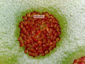
Fern Sorus and Sporangia Movement. Close-up of fern sorus with measurement of 3.82mm. Individual sporangium can be seen in both the photo and video. The video highlights how the sporangia can “open and close.”

Ferns begin to produce spores only once they are fully matured. When a fern’s fronds are totally unfurled, the clusters of sporangia, called sori, can begin to form. The sori tend to be on the underside of the fronds in most species of fern. Some species like those in the Adiantum genus, actually have the sori and indusium, the protective covering, along the edge of the fronds. These are called “marginal indusium.” They are often brown in color, and once they mature they darken and lose their protective cover so that spores can be dispersed. These sori that house the sporangia are actively producing spores during the summer in areas like Ohio. In some tropical locations, the sporangia can be active all year long. Ferns are known as homosporous plants since their spores are all roughly the same size and they all perform the same function (UPenn.edu).

10    11
Marginal Indusium. Marginal indusium as seen on maiden hair ferns (Adiantum sp.).  A younger fern is pictured on the left while an older fern is pictured on the right.

12
Mature and Immature Indusium. The leaf on the left has the more mature indusium than the leaf on the right, as can be told from the darker color of the protective covering.

Relationships Between Ferns and Other Organisms

Some herbivorous vertebrates eat young fiddlehead ferns in the wild. As the fern grows into an adult, it begins to produce toxins in its fronds that discourage animals from eating them. These toxins are still present in the young ferns, but in lesser amounts. Insects like grasshoppers and snails can eat adult ferns on a regular basis despite the increased toxicity. In the time of the dinosaurs, ferns were actually the main food source for the herbivorous sauropods. Today, many vertebrate animal species that eat ferns only eat certain species at certain times of the year (Walker 2010).

Although some young ferns are safe for animal consumption, there are species like bracken ferns that contain toxins called thiaminase and ptaquiloside. These ferns are eaten as fiddleheads by white-tailed deer and eastern cottontail rabbits with little consequence. Sometimes, when vegetation is scarce, some other animals may resort to eating bracken while others avoid it. Livestock, like swine and horses, can ingest bracken and experience thiaminase-mediated syndrome (Veterinary Public Library 2005). Ptaquiloside has been correlated with esophageal or gastric cancer in humans if it is ingested (Stegelmeier 2014).

Despite their miniscule size, spores can also be a food source for animals. The European woodmouse eats the spores of the European fern Culcita macrocarpa, and it is the only small mammal known to do so. The spores are rich in lipids, and they provide a lot of energy in a very small package. The bullfinch and short-tailed bat are the only other two vertebrates known to feed on the spores of a fern (Walker 2010).

Ferns also have a connection with other organisms. A well-known symbiotic relationship of ferns is with mycorrhizal fungi. This relationship is known as mycorrhizas, meaning “fungus-root,” and it is a mutualistic symbiosis between fungus and root systems of plants. Mutualistic symbiosis means that these two organisms live together and each benefit from the other. The evolution of mycorrhizas is thought to have progressed from an endophytic relationship to the ectophytic relationships that we see today. “Endophytic means tendency to grow inward into tissues in fingerlike projections. Ectophytic means tendency to grow outward beyond the surface ipithelium from which it originates” (Brundrett 2002). During the evolution of this relationship, both partners were independent and over time became interdependent due to the exchange of limiting resources between the two (Brundrett 2002).

fern-myco
Mycorrhizal Relationships. The drawing better illustrates mycorrhizal relationships. The thick black line represents the network of hyphae and the blocks represent the cell wall of the plant.

Primitive ferns like the wisk fern (Psilotum)and the horsetail fern (Equisetum) species have subterranean gametophytes that have Vesicular-Arbuscular Mycorrhizae (VAM) associations. VAM means that fungus grows into the root cells of the host to form two kinds of specialized structures: arbuscules and vesicles (McGraw-Hill Dictionary 2003). The arbuscule is an organ in the fungus that absorbs nutrients and vesicles are structures used for storage.  Primitive ferns actually lack arbuscules, but are still considered to have VAM associations. Studies have shown that the majority of complex ferns also have these VAM associations. Most ferns have fine roots with long root hair-like structures, suggesting they have mycorrhizal association that helps with function. Leptosporangiate ferns are considered to be the least dependent on mycorrhizas (Brundrett 2008).

Diversity of Ferns and Their Uses

13
Linear Sori. The birds nest fern (Asplenium nidus) is part of the family aspleniaceae, in which all members display linear sori.  The sori hold the spores and are the brown linear lines in this image.

15
Specialized Rhizomes. Rabbit’s foot fern (Davallia solida) is known for its thick, fuzzy, rhizomes that creep along the ground or down the side of the pot when placed in a hanging pot.

There are many types of ferns that make up the vast diversity of the taxa such as: bird’s nest fern, boston fern, button fern, rabbit’s foot fern, holly fern, staghorn fern and ostrich fern.  They are all unique because of the different ways some of their structures are modified.  For example some ferns like the rabbit’s foot fern shown above have modified their rhizomes to creep along the ground whereas some ferns rhizomes are in the ground.  The bird’s nest fern, also shown above, has modified linear sori.  The different ways the sori are positioned on the leaf is another example of fern diversity.

14
One Leaf or Many Leaflets? The golden polypody fern (Phlebodium aureum) shows how not all ferns have leaflets.  This fern technically doesn’t have leaflets, because the fronds are not separated near the stem, making this all one leaf.

Hokkaido, an island in northern Japan, has approximately the same number of species of ferns as North Eastern America. About 40% of these ferns are considered sister species due to their similar characteristics. Being on opposite sides of the globe it is strange to see species that are so similar without a clear pattern of distribution but such is the case with these ferns. This could be in part due to the similar climates in both regions giving rise to similar evolutionary characteristics (Moran, 2004).

Ferns, unlike some other plants, are not of major economic importance to humans today even though they are a popular horticultural plant. Since they were so prevalent during the Carboniferous Period, the fossils of ancient ferns have actually contributed to the world’s supply of fossil fuels (science.Jrank.org). Our modern ferns still have uses for us today. Many people will grow ferns as ornamental plants and some will search for certain fiddlehead species like the ostrich fern (Matteuccia struthiopteris) for a spring-time treat to put in salads or used as a steamed vegetable.

Tree ferns of the families  Dicksoniaceae and Cyatheaceae are also of commercial importance. The trunks of the tree ferns can be used in construction work. The root system of the tree fern also is widely used as a substrate to grow ornamental orchids. Stalks and buds of these ferns can also be made into packing material for pillows and mattresses. This led to the destruction of many populations of tree ferns in tropical locations (Reference.com).

161819  

Tree ferns.  The first photo on the left is Japanese wood ferns (Dryopteris sieboldii).  The middle photo is a Hawaiian tree fern (Cibotium glaucum). The right photo is a giant fern (Angiopteris evecta).  Although they are known as tree ferns they are not true trees and the area that looks like a trunk is really just made out of leaf bases.

Different fern species are also used in many Eastern countries as some form of medicine. The study of traditions and uses of plants as medicine in different cultures is known as ethnobotany (Dictionary.com). In the Himalayas, rattlesnake fern (Botrychium virginianum) is turned into a lotion that is spread onto sores, cuts, bruises, and burns. The rhizomes and petioles of the Indian button fern (Tectaria macrodonta) are used in India to relieve constipation (OpenSIU).

Ferns have also been shown to help remove heavy metals, like arsenic, from soil. Arsenic can enter the environment through human activities and is commonly found in pesticide, dyes, and wood preservatives. Arsenic in high doses can be poisonous to people, animals, and many plants (USF.edu). High arsenic levels in soils have become a problem since arsenic-related health problems are increasing all over the world. In 2001, it was discovered that the Chinese brake fern (Pteris vittata) grows well in arsenic-contaminated soil. It accumulates excess arsenic in the fronds and is an environmentally-friendly and cost-effective solution to arsenic-rich soil. Many studies have been done to observe arsenic uptake into the fronds, and the effects that this process may have to other organisms that eat fern fronds (Fayiga et al. 2004).

Why Are Ferns Important?


Ferns have contributed a lot to earth’s natural history. They have provided an important evolutionary stepping stone for land plants as we know them today: vascular tissue. Without plant vascular tissue, we could not have the giant sequoias of California or the orchards of fruit trees that we cultivate. Their fossils have gone on to make important fossil fuels like coal and natural gas. Ferns can also uptake heavy metals from the soil and aid in healing contaminated environments.

Ferns have many uses today that people don’t really think about as well. Some animals use ferns as a source of food. Humans can use ferns as ornaments, or use the tree ferns to help build structures. Even though they are not as charismatic as other vascular plants, they are very interesting and provide us with a glimpse to the past. Hopefully the next time you look at a fern, you will remember everything that they do.

Works Cited:

American Fern Society. (2006). A Brief Introduction to Ferns. Retrieved from: http://amerfernsoc.org/lernfrnl.html

Australian National Botanical Gardens. (1998, July). Ferns as ecological indicators. Retrieved from: http://www.home.aone.net.au/~byzantium/ferns/ecol.html

Australian National Botanical Gardens. (1998, July). About ferns. Retrived from: http://www.home.aone.net.au/~byzantium/ferns/about.html

Brundrett, Mark C. “Coevolution of roots and mycorrhizas of land plants.” New phytologist 154.2 (2002): 275-304.

Brundrett, M. (2008). Mycorrhizal associations: the web resource. Retrieved from: http://mycorrhizas.info/evol.html

Ecological niche. Dictionary.com Unabridged. Retrieved March 29, 2015, from Dictionary.com website: dictionary.reference.com/browse/ecological+niche

Ensminger, PA. (2009). Ferns – importance to humans. Retrieved from: http://science.jrank.org/pages/2689/Ferns-Importance-humans.html

ethnobotany. Dictionary.com Unabridged. Retrieved March 29, 2015 from: dictionary.reference.com/browse/ethnobotany

Fayiga, Abioye O et al. “Effects of heavy metals on growth and arsenic accumulation in the arsenic hyperaccumulator Pteris vittata L.” Environmental pollution 132.2 (2004): 289-296

McGraw-Hill Dictionary of Scientific & Technical Terms, 6E. (2003). Vesicular arbuscular mycorrhizal fungi. Retrieved March 29 2015 from: http://encyclopedia2.thefreedictionary.com/vesicular-arbuscular+mycorrhizal+fungi

Merriam-Webster Dictionary. (2003). Epiphytic. Retrieved from: www.merriam-webster.com/dictionary/epiphytic

Moran, Robin C. (2004). A Natural History of Ferns. Oregon. Timber Press.

Srivastava, Kamini. (2007) “Importance of ferns in human medicine.” Ethnobotanical Leaflets 2007.1: 26.

Stegelmeier, BL. (2014, Feb) Overview of bracken fern poisoning. Retrieved from: http://www.merckmanuals.com/vet/toxicology/bracken_fern_poisoning/overview_of_bracken_fern_poisoning.html#v11577387

The encyclopedic entry of fern. (2008). Retrieved March 30, 2015 from: http://www.reference.com/browse/fern

Veterinary Public Library. (2005). Bracken Fern or Brake Fern (Pteridium aquilinum). Retrieved from: http://www.library.illinois.edu/vex/toxic/bracken/bracken.htm

Walker, M. (2010, Feb 19) A mouse that eats ferns like a dinosaur. Retrieved from: http://news.bbc.co.uk/earth/hi/earth_news/newsid_8523000/8523825.stm

Part 2

Lifecycle & Ecology

Angiosperms are considered to be one of the greatest examples of symbionts in nature and due to their many mutualistic relationships with pollinators, fungi, herbivores and others. They can be found in almost any environment, so long as there is sunlight, some form of water, and a way to spread their offspring. There are four major events that make up flowering plant reproduction; pollen development, egg development, pollination and fertilization. Angiosperms produce two types of spores; microspores which lead to the generation of pollen and megaspores which form the structure that houses female gametophytes (Boundless, 2014).

Pollen develops inside the stamen. Inside the anther of an angiosperm lie the diploid microspores. These microspores undergo meiosis to become haploid microspores. Haploid microspores then undergo mitosis to develop into pollen grains. Pollen forms from the male gametophyte in flowering plants.  The gametophyte consists of two cell types: tube cells to aid in fertilization and generative cells which generate sperm cells (Taiz, 2006).

Egg development occurs inside the carpel. Inside of the ovaries are egg producing structures known as ovules. Inside of an ovule are diploid cells. Similar to the production of pollen, these diploid cells divide via meiosis to become haploid cells that are the megaspores. These spores then go through three rounds of mitosis forming seven cells. One of the cells has two nuclei, called the endosperm and another of the cells becomes the egg. The remaining five cells are not used in reproduction (Boundless, 2014).

Once both eggs and pollen development have taken place they are ready for pollination. Pollination can occur in many ways; two major forms are wind and water dispersal. Unique to angiosperms is the use of pollinators such as birds and bees.  The last step in flowering plant reproduction is fertilization. For fertilization to occur the tube cell of the male gametophyte creates a tube to the ovule (Taiz,  2006). The generative cell uses this tube to send sperm down to the ovule so fertilization can occur. One sperm will fuse with the egg forming a zygote the other fuses with the endosperm forming a triploid endosperm cell. This process, known as double fertilization, is unique to angiosperms (Derksen, 2013). The endosperm later develops into nutrient tissue while the zygote divides by mitosis, developing into an embryo which grows into a mature plant. Angiosperms spend most of there life in the adult stage known as a sporophyte. When we see trees, grass, flowers, vegetables in a garden we are seeing sporophytes! Angiosperms are very important due to their abundance and impact on almost every habitat on earth. Due to their diverse morphology they can range from the small to massive, aquatic to mountainous, grass to trees and everything in between.

Evolutionary History

The Amorphophallus titanum, or Corpse flower is one of the most bizarre and oldest ancestors of modern angiosperms. Photo taken by Nick White

The Amorphophallus titanum, or Corpse flower is one of the most bizarre and oldest ancestors of modern angiosperms. Photo taken by Nick White

Not surprisingly, angiosperms are the most commonly found type of land plant. Angiosperms evolved in the Cretaceous era, around the same time as many groups of modern insects. Many of these insects acted as pollinators that drove the evolution of both angiosperms and the insects themselves. (Soltis 2005). Due to the availability of pollinators (insects occupy nearly every environment on the planet) it has allowed angiosperms to become the most numerous plant found on land. This relationship is considered one of the greatest examples of symbioses in nature due to their many mutualistic relationship with pollinators, fungi, herbivores and others. They can be found in almost any environment, so long as there is sunlight, some form of water, and a way to spread their offspring.With co-evolution, these two species have been able to occupy places that few other species previously could, changing the habitats of the entire planet.(Lerner 2008)

Unlike many land plants, angiosperms did not evolve from gymnosperms. It is unclear what type of plants gave rise to angiosperms. (Angiosperms 2014) Some scientists believe that a group of plants known as “seed ferns” ,or pteridosperms, may have been the progenitor of the angiosperms. These “seed ferns” were around for many millions of years before angiosperms and yet have similar traits like seed-bearing capsules and specialized organs that produced pollen. While we are still not exactly sure how ancient angiosperms may have come about, we have an idea of what these ancestors may have looked like. They were likely small with small flowers. The flowers were probably green and not at all like the flowers we are used to as their sepals and petals would not be separated or distinguishable (Angiosperms 2014). And while the exact way that angiosperms evolved to what we know today is still unclear, their impact on our world today is obvious.

Porifera Part Two

Porifera Life Cycle

The life cycle of a sponge is a relatively simple one. Sponges can reproduce sexually and asexually. There are many sponge species in which each sponge is considered male and female. When it comes to sexual reproduction, a sponge can play either role. The male sponge releases sperm into the water which travels towards and enters the female sponge. After fertilization occurs, a larvae is released from the female sponge into the water. The larvae floats around for several days until it can find a suitable substrate to stick to. At this point, the larvae will begin to grow into an adult sponge. Sponges become more diverse when different sponge species reproduce with one another (Myers, 2001). The sexual life cycle is depicted below in Figure 1.

When sponges go through asexual reproduction, it is by a system called budding. This occurs when a small piece of the sponge is broken off and is able to grow into a whole new sponge. Like in sexual reproduction, this small piece of the sponge must find a substrate to cling to in order to grow into an adult sponge (Myers, 2001).

pic 1

Figure 1. The life cycle of a sexually reproducing sponge. Hermaphroditic sponges acting as “male” (A) release sperm into the water. A sponge acting as “female” (B) receive sperm through their pores. Sperm is directed into the sponge wall where the egg is located (C). Fertilization occurs and a zygote forms (D). The zygote is released into the water and uses its cilia to swim to a new location (E). Once the zygote settles into habitable substrate, it begins to grow into a mature sponge (F). Drawing by Sarah Petersen. Information from Myers (2001).


Sponges and their Associations with Other Organisms

Sponges make up an important component of coral reefs and filter the surrounding water and cycle nutrients (Hultgren, 2014). Sponges have a wide range of associations with other organisms, which can include facilitating primary production, providing a habitat for another organism, or even providing protection to organisms from predation. Sponges interact with a wide range of organisms, so it is sometimes difficult to understand the role sponges play in these relationships (Bell, 2008).

To facilitate primary production, sponges associate themselves with photosynthetic organisms. A review paper by Bell (2008) mentions that these photosynthetic relationships contribute between 48 and 80% of the sponge’s energy requirements and around 10% of the reef’s productivity. This paper also mentions that the role of sponges as primary producers may only be important for nutrient-poor waters, such as those found in the tropics. Sponges are also involved in secondary production because other organisms such as fish, crustaceans, and molluscs consume them. These predators vary over the differing ecosystems that sponges are found in (tropical, temperate, polar, etc.). Since sponges harbor photosynthetic organisms, the sponges being eaten by predators could be seen as a herbivorous interaction because the photosynthetic organisms may be of greater nutritional value than the actual sponge itself (Bell, 2008).

Sponges also provide microhabitats for smaller species. Costs and benefits to each organism in the relationship are not well studied. In an experiment by Hultgren (2014), the relationship between the Synalpheus species of snapping shrimp and the marine sponges they inhabit. It was found that the shrimp had varying effects on the sponges. When predators were present, the shrimp had positive effects on the sponges and negative effects during periods when the sponge was actively growing. These negative effects were likely the shrimp consuming the sponge as it grew. This study suggested a future study in which abiotic and biotic stressors should be manipulated to see if the relationship between shrimp and sponge changes. Sponges also interact with other organisms such as bivalves. When bivalves had sponges living on their valves, their risk of predation by starfish was reduced. The sponges benefit from the association by having an increased feeding efficiency. Sponges are also associated with crabs, which have been observed to use the sponges to cover their bodies as a form of camouflage (Bell, 2008).


Where Can Poriferans be Found?

Sponges have a global distribution that encompasses polar and tropical latitudes alike.  They can be found from deep depths in the ocean to shallow rock pools.  Poriferans have been known to occupy habitats that include marine thermal vents as well as freezing arctic waters.  While many may associate sponges with a solely marine habitat, a portion of the phylum occupies freshwater as well such as the preserved specimen pictured below, found near a hatchery in New York.  These freshwater sponges are represented by 219 out of the 15,000 species of Poriferans, all belonging to the suborder Spongillina of the class Demospongiae.  While the diversity of freshwater sponges may be limited in comparison to their marine relatives, their abundance within their freshwater habitats is often greater.  What is most amazing about freshwater sponge diversity is the specificity of the taxa found occupying a body of water.  Around 47% of freshwater sponges originated from the body of water they inhabit, and new species discoveries can often been associated with new genre as well (Manconi and Bronzanto, 2008).

pic 2

Figure 2. A preserved freshwater sponge from New York. Picture taken by Christine Koporc at the OSU Museum of Biological Diversity.

As mentioned earlier, sponge fossils have been found dating back to the Precambrian, where poriferan remnants have been identified in marine strata (Porifera: Fossil Record, 2006).  Colonization of freshwater by sponges has been dated back to the Mesozoic period, which occurred some 220 million years ago.  This theoretical date is based off of structures found in the oldest known freshwater sponge fossils, called gemmae.  These are highly conserved features within Spongellina, and act as asexual propagules of the sponge which can become dormant if environmental conditions are not favorable.  This is a feature that has been associated with the ability of sponges to occupy some of the unique inland habitats they are found in (Manconi and Bronzanto, 2008).  Until relatively recently in Earth’s geological time scale, sponges have constituted a large part of the framework of oceanic reefs.  Today, sponges still remain an important part of reef communities, however do not contribute nearly as much to mass as corals (Porifera: Fossil Record, 2006).


Diversity in Sponges

There are many times when it is difficult to differentiate between two sponge species. On more than one occasion, two sponge species were thought to be one until scientists took their observations of the sponges one step further and realized that there was a difference between the two. There are various ways in which sponge species can be distinguished from one another. Some are seen as a food source to certain animal species while other very similar sponges are not, such as seen in fire sponges (Tedania ignis) and volcano sponges (Tedania klausi). These two sponge species used to be seen as one diverse species, fire sponges, until scientists performed feeding choice experiments as well as a morphological and molecular study and determined that they were not the same species at all. Volcano sponges are eaten by sea stars, while fire sponges are not, leaving the species to live in very different habitats. These two species were also found to have a difference in their susceptibility to disease and ability to withstand a wide change in temperature and salinity. Molecular markers also helped scientists to see the difference between the sponge species Scopalina blanensis and Scopalina lophyropoda. S. blanensis responds positively to seasonal environmental changes in temperature and food availability, while S. lophyropoda responds similarly throughout these environmental changes (Wulff, 2012).

Differences in sponge species can be seen in many different aspects of the sponge and its life. They can be based upon the role that a sponge takes in its community, their association with other species, morphological (physical) characteristics, and their vulnerability to hazards. An example of the morphological differences between sponges can be seen below in Figures 3 and 4. Some of these contrasting features can be determined through observation, while others require the use of experiments (Wulff, 2012).

pic 3

Figure 3

pic 4

Figure 4


Figures 3 and 4. The physical diversity of sponges is clearly seen between these two dried marine sponge specimens. Pictures taken by Christine Koporc at the OSU Museum of Biological Diversity.


Freshwater and Saltwater Sponges

Sponges live in a wide variety of ecosystems. Sponges are such simple organisms that they have been able to adapt to many different environments, which is why they are able to be found in nearly every type of body of water. They are found in the deep sea, in coral reefs, near hydrothermal vents (which are 3,000 to 7,000 feet below the water’s surface), and in various freshwater environments (Masters, n.d.). While most live in marine environments, such as oceans, there are still some that live in freshwater environments, such as lakes, ponds, and streams. Around 200 of the 15,000 known species of sponges live in freshwater environments (Skelton & Strand, 2012). The important functions of marine sponges have been determined by their impact on substrate (reef creation and erosion of hard oceanic substrate), their coupling with other organisms living at the bottom of the sea (their role in the carbon cycle, silicon cycle, nitrogen cycle, and oxygen depletion), and their interactions with other organisms (Bell, 2008). Much less is known about freshwater sponges and the roles that they play in their ecosystems, though it is believed that their roles will be very similar to those that are seen in marine sponges (Skelton & Strand, 2012). Even with this limited amount of information for their functional roles, freshwater sponges are found to be very hardy creatures that can withstand a wide variety of situations, such as drought, chemical pollution, and fluctuations in water flow, pH, and temperature (Masters, n.d.).


How sponges are used today

Sea sponges have vast economic importance. The types used are mainly demospongiae because they possess spongin which is the flexible skeleton like structure of the sponge.

Video 1. Student demonstrates the flexibility of the spongin using a dissecting microscope.


The most popular sponge used is the Wool sponge; it is the softest and most durable sponge. Firstly, they are used in industry. There is a city in Florida called Tarpon Springs located in the Gulf that is the acclaimed Sponge Capital of the World. They harvest and export a majority of the worlds sponges. Before World War II, Florida produced 600,000 pounds that is 7,800,000 individual sponges on average for human use. In recent years they produce around 70,000 pounds, that is 910,000 individual sponges on average (Stevely & Sweat, 2015).

Figure 5. Freshly harvested wool sponge. Photo taken by Christine Koporc at the Sponge Docks in Tarpon Springs, Florida.

Sea sponges are very popular in the health and beauty field. They can be used for cleaning an array of surfaces and have better water retention than that of the artificial sponge. Most popular uses include car care, household cleaning, makeup application and removal, skin exfoliant for when bathing, and personal care. If taken care of properly, they can last years on end where an artificial sponge can fall apart and be riddled with bacteria after months of use.

Figure 6. Above is an example of how a wool sponge is implemented in a bar of soap for health/beauty purposes. Photo taken by Christine Koporc in North Port, Florida.

Figure 6. Above is an example of how a wool sponge is implemented in a bar of soap for health/beauty purposes. Photo taken by Christine Koporc in North Port, Florida.

Other organisms use sponges

Dolphins will use marine sponge to protect themselves while searching for food. They will grab a sponge from the seafloor and fit it around their beak to protect it from chunks of coral or rock that could hurt them. It is hypothesized that they hunt the bottom dwelling fish instead of the ones out in the open ocean because the bottom dwelling fish are more nutritious (Morell, 2011). Also, a variety of microorganisms, worms, crabs and shrimp will inhabit the cavities in the sponges. Sponges also serve as a protection mechanism for scallops. The sponges will attach and live on the shell and protect it from organisms such as starfish which can damage it (Bean-Mellinger, 2015).

Human Impact

Sponges have been harvested since the 1800’s because they are beneficial and durable for many uses. They are a huge industry in Florida. The regeneration of the sponges that are harvested is important for the health of the ecosystem; there are now certain parts of Florida where harvesting sponges is illegal. In the beginning sponges were harvested using the hook method. This entails a diver using a pronged hook to grab the sponge then rip it free of its base. It was later discovered that the hook method inhibits the chance of that sponge fully regenerating. It is now a law that sponges have to be harvested using the cutting method, making sure to leave enough left at the base of the sponge for proper re-growth. This entails using a knife to cleanly cut the sponge away. Doing this brings the chance of survival for the sponge to 71% versus 41% for hooked ones (Stevely & Sweat, 2015).

Figure 7. Wool sponge laying on its side. Left side of the sponge is the bottom; it can be seen it is flat because it has been cut, not torn from its base. Photo taken by Christine Koporc in North Port, Florida.

Conclusion

It’s now easy to see why we can appreciate phylum porifera and its place in basal Metazoa.  What may represent one of the first multicellular organisms that successfully survived and diversified to current day is also a reminder of Metazoa’s more humble beginnings in evolutionary history.  While these hermaphroditic organisms may not have organized tissue, they certainly have specialized cells and organelles to carry out the functions that would otherwise be performed by more complex structures in higher animals. Though their mobility is limited to larval stages, gametes, and the occasional passive gemmae in freshwater specimens, these organisms have also managed to occupy an incredible diversity of habitats, all at varying depths, latitudes and longitudes.

Sponges have had a large impact on their environment as well.  This includes not only their primary role in reef structure throughout geological history, but direct use by other animals.  Animals like humans, who rely on marine sponges in particular as an industry, can also have a significant influence on their survival through overharvesting.  Poriferans are certainly amazing creatures, and their ancient lineage in conjunction with their diversity in taxa, habitat, chemical production, and ecological utility represent the merit in appreciation of these organisms.


Literature cited

Bean-Mellinger, B. (2015). Relationship Between Scallops and Sponges. Animals. Retrieved from http://animals.pawnation.com/relationship-between-scallops-sponges-9329.html.

Bell, J. J. (2008). The Functional Roles of Marine Sponges. Estaurine, Coastal and Shelf Science, 79(3), 341–353.

Hultgren, K.M. (2014). Variable effects of symbiotic snapping shrimps on their sponge hosts. Marine Biology 161, 1217-1227.

Manconi, R., Masters, R. (2008). Global Diversity of Sponges (Porifera: Spongillina) in freshwater. Hydrobiologia, 595(1), 27-33.

Masters, M. (n.d.). Habitats of Sea Sponges. Retrieved from http://animals.pawnation.com/habitats-sea-sponges-2396.html.

Morell, V. (2011, July). Why Dolphins Wear Sponges. Science. Retrieved from http://news.sciencemag.org/environment/2011/07/why-dolphins-wear-sponges.

Myers, P. (2001). Porifera Sponges. Retrieved from http://animaldiversity.org/accounts/Porifera/.

Porifera: Fossil Record. (2006). Retrieved March 28, 2015, from http://www.ucmp.berkeley.edu/porifera/poriferasy.html.

Skelton, J., & Strand, M. (2012). Trophic ecology of a freshwater sponge (Spongilla lacustris) revealed by stable isotope analysis. Hydrobiologia, 709(1).

Stevely, J., & Sweat, D. (2015). Florida’s Marine Sponges: Exploring the Potential and Protecting the Resource. Retrieved March 28, 2015, from http://edis.ifas.ufl.edu/sg095.

Wulff, J. (2012). Ecological Interactions and the Distribution, Abundance, and Diversity of Sponges. In Advances in Marine Biology (pp. 273–344).


Hexapods

Introduction to Hexapods


Welcome to the Hexapod Blog! What really is the story behind those creepy crawly critters? They may not be as disgusting and scary as you think! Hexapods have a rich evolutionary history and are one of the most diverse groups of animals on earth. Follow us as we dive into hexapods, and learn all about their biology. Maybe before you squish one under your shoe, you will remember how awesome and beneficial they can be!


We have everything you need to know about hexapods right here! First, we provide some history. How long ago did they first appear? What does their family tree look like? We have all heard of dinosaur fossils, but what about hexapod fossils? In fact, hexapods do have their own fossil record which we also discuss. Second, we dive into what makes a hexapod a hexapod. The insect body plan, wings, wing folding, and complete metamorphosis are the main key evolutionary innovations we focus on. For instance, the mealworm beetle, Tenebrio molitor, undergoes complete metamorphosis changing from a mealworm larvae, pupa, and finally an adult beetle. The field cricket, Gryllus pennsylvanicus, is a good example of the insect body plan. They have a clear head, thorax, and abdomen. Of course it would not be a hexapod if it did not have six legs! The Madagascar hissing cockroach, Gromphadorhina portentosa, is one such critter having three sets of legs. Find all this and more in the Hexapod Blog!

Phylogeny


There are four different supergroups that help to classify Eukaryotes and each of their lineages. These groups are known as the Chromalveolates, the Excavates, the Archaeplastida, and the Unikonts. Hexapods can be found later down the Unikont lineage (Tree of Life Web Project, 2002).


The Unikonts split into various lineages, including Fungi, Amoebozoa, and others. However, Hexapoda is found in the Animal lineage. The lineage of Hexapoda continues to another group deemed Bilateria that includes most groups that are considered to be animals (Tree of Life Web Project, 2002). Scientists have determined that there are two separate lineages that make up Bilateria, which are Deuterostomia and Protostomia. Deuterostomes and Protostomes are defined by their embryonic development. Deuterostomes develop their anus before their mouths, and Protostomes develop their anus after their mouths. Scientists now recognize two lineages within Protostomia that are Lophotrochozoa and Ecdysozoa (Halanych et. al, 1995). Hexapods can be found in the Ecdysozoan or molting insect lineage under the phylum Arthropoda (Tree of Life Web Project, 2002).


Hexapoda are traditionally shown to be monophyletic or descended from one evolutionary group. They are one of the most diverse groups, with over 750,000 species described so far (Tree of Life Web Project, 2002). Recent evidence suggests that the closest relatives to Hexapoda are Crustacea (Giribet et. al, 2001). While Crustacea are dominant in aquatic environments, Hexapoda are dominant on the land. The Hexapods split into lineages that include Diplura, Insecta or the true insects, Protura, and Collembola or the springtails (Tree of Life Web Project, 2002). Of each of these groups, Diplura is seen as the most unstable and it’s place in the phylogeny of Hexapoda changes often (Tree of Life Web Project, 2002). A final taxonomy of insects would be ordered as Eukaryotes, Unikonts, Animals, Bilateria, Ecdysozoa, Arthropoda, Hexapoda.

IMG_0040

Phylogenetic tree of Hexapoda. Created using Class Notes and J.E. Blair, 2009. Created by Daniel Pinto


Fossil Evidence


Hexapods are one of the earliest known lineages of terrestrial based insects, dating all the way back to the Early Devonian period about 412 million years ago or possibly even to the Late Silurian period (Misof et. al, 2014). These early fossils are that of Rhynies (Rhyniognatha hirsti), or springtails (Collembola). Some basal insect fossils could date to 379 million years ago, however the fossil evidence is sparse and therefore scientists are unable to say definitively what lineage these fossils are from. However, fossils of other early insects include those of bristletails (Archaeognatha) that date to about 390 million years ago (Engel et. al, 2004). Most of this evidence suggests that insects were one of the first terrestrial organisms and were major selectors on land based plant life (Engel et. al, 2004). Fossil evidence of key structures have helped scientists to determine if such structures allowed for the massive speciation of the Hexapod lineage (Nicholson et. al, 2014). The fossil record can be a good way to show early existence of particular groups and can help to infer earlier origins if some groups are more diverse or abundant than others (Thomas & Ware, 2011). However, the fossil record is not as complete for certain groups of Hexapods, thus making it not as reliable as other dating methods (Thomas & Ware, 2011).

Molecular Clock


Unlike the fossil record, molecular clock evidence can be seen as more reliable in some cases. Using genomes of living Hexapods, estimates for divergence dates of certain groups can be found even in the midst of a sparse fossil record (Thomas & Ware, 2011). In a study on the evolutionary history of insects, molecular clock data led scientists to conclude that Hexapods may have originated earlier than the Silurian period in the Cambrian or Early Ordovician periods (Misof et. al, 2014). These results have been controversial, as there are little to no actual fossils of Hexapods from the Cambrian to the Silurian periods (Misof et. al, 2014). Indeed, some scientists have hypothesized that Hexapod evolution happened drastically earlier than paleontological evidence based on molecular evidence (Thomas & Ware, 2011).


Though there are benefits to using molecular clock data, there are also drawbacks as well. Using molecular clock data can help to fill gaps in the fossil record, but data can only be obtained from living or recently extinct species. This is due to the fact that molecular clock data relies on using DNA sequencing to infer genetic changes over time, but genetic material cannot be obtained from ancient species (Thomas & Ware, 2011). Despite these limitations, molecular clock data has supported the idea of Hexapods being a monophyletic group, and even helped to confirm previous hypotheses about the close relation between Hexapoda and Crustacea (Misof et. al, 2014).

Key Evolutionary Innovations of Hexapods


Hexapods are one of the most diverse classes in the animal kingdom. In fact, Hexapods alone make up over half of all recorded species. With such diversity and success, you can bet there a few innovations that have warranted their success.


  1. Hexapod bauplan: The importance of the bauplan, or body plan, of Hexapods cannot be overlooked when it comes to contributing to their success. Hexapods exhibit metameric, or repeated, segmentation and have one pair of appendages per segment. These segments are organized into three tagmata, or specialized segments: the head, thorax, and abdomen. Their appendages are jointed, which aids in walking, swimming, and feeding, similar to the joints in our bodies. Hexapods also have exoskeletons, meaning their skeletons are on the outside of their bodies, providing them with protection (Nicholson et. al, 2014).

Schneider_Cricket_photo_1

Field Cricket, example of insect bauplan


  1. Wings: Hexapods are thought to have begun flying sometime in the Carboniferous Period, about 350 million years ago. While it is not fully understood why wings developed, the hexapod wing and the flight capabilities it provides are extremely advantageous. Hexapod wings are paired, and the pairs are generally referred to as the fore- and the hind-wings. They are heavily veined, and are surrounded by a cuticle for rigidity and protection. Most Hexapods have their fore- and hind-wings coupled, but dragonflies do not; they can move each of their wings completely independently of the others, allowing them to perform amazing flight maneuvers (Kesel, 2000). The benefits of flight are many, and wings have no doubt lent to the overwhelming success of Hexapods and other insects (Yanoviak et. al, 2009).


IMG_0037

Cicada exhibiting folded-over, heavily veined wings.

  1. Metamorphosis: There are two types of metamorphosis, hemimetabolism and holometabolism. Hemimetabolism, known as incomplete metamorphosis, characterizes Hexapods that go through a series of molts, shedding the exoskeleton that has become too small for them. Holometabolism, or complete metamorphosis, is a four stage cycle in which the organism goes from egg to larva to pupa to adult. Hexapods that undergo hemimetabolism typically resemble the adult stage as nymphs, but holometabolous Hexapods do not. A unique advantage to metamorphosis is that it allows them to inhabit different niches throughout the course of their life (Class Notes). For example, a grounded caterpillar will not occupy the same niche when it becomes a winged adult butterfly.  In addition, it has been suggested that holometabolous Hexapods enjoy lower extinction rates than other groups (Nicholson et. al, 2014).


  1.  Sensory systems: Hexapods have acute, highly developed senses. They have compound eyes, and each eyeball is comprised of tiny units called ommatidia. Each ommatidium contains its own lens. In addition, most Hexapods also have simple eyes, or ocelli, to further their sense of sight (Mayer, 2006). Many Hexapods, such as wasps and mantids, have two large compound eyes and three smaller ocelli on top of their heads. In addition to their developed eyesight, Hexapods were among the first creatures to sense sounds. Most sound is produced through repeated stimulation of appendages. Some species of moths can even hear ultrasound, helping them avoid predation by bats (Kay, 1969).

A review of key evolutionary innovations of Hexapods, including metamorphosis, wing folding, and body segmentation.

References

Engel, M. S., & Grimaldi, D. A. (January 01, 2004). New light shed on the oldest insect. Nature, 427, 6975, 627-30.


Giribet, G., Edgecombe, G. D., & Wheeler, W. C. (January 01, 2001). Arthropod phylogeny based on eight molecular loci and morphology. Nature, 413, 6852, 157-61.


Halanych, K. M., Bacheller, J. D., Aguinaldo, A. M., Liva, S. M., Hillis, D. M., & Lake, J. A. (January 01, 1995). Evidence from 18S ribosomal DNA that the lophophorates are protostome animals. Science (New York, N.Y.), 267, 5204, 1641-3.


J.E. Blair. Animals (Metazoa). Pg. 223-230 in The Timetree of Life, S.B. Hedges and S. Kumar, Eds. (Oxford University Press, 2009).


Kay, Robert E. (1969). “Acoustic signalling and its possible relationship to assembling and navigation in the moth, Heliothis zea”. Journal of Insect Physiology 15 (6): 989–1001.


Kesel, A.B. (2000) Aerodynamic characteristics of dragonfly wing sections compared with technical aerofoils. J Exp Biol 203: 3125–3135.


Mayer, G. (2006), “Structure and development of onychophoran eyes: What is the ancestral visual organ in arthropods?”, Arthropod Structure and Development 35 (4): 231–245.


Misof, B., Liu, S., Meusemann, K., Peters, R. S., Donath, A., Mayer, C., Frandsen, P. B., … Zhou, X. (November 06, 2014). Phylogenomics resolves the timing and pattern of insect evolution. Science, 346, 6210, 763-767.


Nicholson, D.B., Ross, A.J., Mayhew, P.J. Fossil evidence for key innovations in the evolution of insect diversity. Proc. R. Soc. B: 2014; 282(1803).


Thomas, J. A., & Ware, J. L. (January 01, 2011). Molecular and Fossil Dating: A Compatible Match?. Entomologica Americana, 117, 1, 1-8.


Tree of Life Web Project. 2002. Hexapoda. Insects, springtails, diplurans, and proturans. Version 01 January 2002 (under construction). http://tolweb.org/Hexapoda/2528/2002.01.01 in The Tree of Life Web Project, http://tolweb.org/
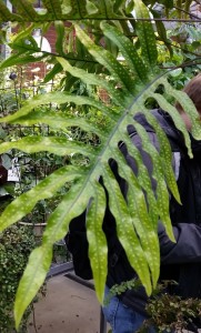
 

One Shell of a Phylum

One Shell of a Phylum


Consisting of more than 85,000 extant species, Mollusca is the second biggest phylum in the animal kingdom, second to only Arthropods. Ten total classes of Molluscs have existed throughout evolutionary history, but only eight of these classes exist today. The word Mollusc was derived from the Modern Latin term mollusca which meant “thin-shelled”. According to Nordsieck (2011) this term originated from the Latin term molluscus, meaning “soft”. These terms were actually initially used to describe many soft-bodied invertebrates that do not fall under this Phylum, including brachiopods, bryozoans and tunicates.


One of the coolest things about molluscs is that their range of adaptations is almost limitless. Most Molluscs have a shell (Fig. 1), a rasping tongue known as the radula, and a foot. What is so fascinating about these traits is that even though they are for the most part, very similar, they give rise to an extremely diverse array of functions that have allowed molluscs to thrive! The molluscs’ reach into so many different areas that its makes other phylums kind of jealous. They inhabit freshwater, marine, and terrestrial environments, utilize various food sources, and move around in very different ways.

Speaking of moving around, Molluscs locomotion is extremely diverse. Modes of movement vary greatly from slow crawling, zipping around through the water, or simply staying still. Some other interesting characteristics of Molluscs include: bilateral symmetry, a body with more than two cell layers, tissues and organs (Nordsieck, 2011).They do not have body cavities, but they do posses a gut with a mouth and an anus. Most have an open-circulatory system with a heart and an aorta, and do gas exchange through organs called ctenidial gills. They reproduce sexually, which can be external or internal, and can even be hermaphroditic!

1 - Ondrejech_Andy_Shell_Variance_Photo

Fig 1 – Some of the largest shells found in each class discussed, and an important shared trait.


We chose to present the European Squid (Loligo vulgaris) representing the Cephalopods (squids, octopuses, nautiluses, cuttlefish),  the Brown Garden Snail (Cornu aspersum) representing the Gastropods (snails, slugs, sea slugs, limpets), and the Blue Mussel (Mytilus edulis, seen in the photo on the right) representing the Bivalves (mussels, clams, oysters, and scallops). These were selected as they are common enough to be found locally allowing us a chance to observe them first hand and collect media on them. Because they are local molluscs, they are more familiar to the general public and are therefore, great candidates to represent the three classes of molluscs that we will sample from for this blog.
In The Beginning…


Fossil evidence shows us that molluscs appeared early in the Cambrian period (about 550 to 580 million years ago) as organisms that crawled along the ocean floor. According to Nordsieck (2011) these fossil records help to explain the division of early molluscs body plans into a soft ventral side used for locomotion (the foot), and an armored dorsal side exposed to the environment. Originally, the dorsal side was protected by a thick tissue layer instead of a shell in order to protect their organs. This was to become the mantle, and can be found in all molluscs. Over time, the mantle developed which, according to Sigwart and Sutton (2007) is a hard horn-like cuticle material that is partly made of the calcium carbonate found in the molluscs’ food sources. This rendered it additionally resistant. Eventually, the tried and true shell developed! While some molluscs developed overlapping shell plates to allow flexibility, others had shell plates that fused together and sacrificed mobility for protection. This one-part shell adaptation proved so successful that molluscs still have it, and it has allowed molluscs to experience a wide range of diversity. For instance, according to the University of Cambridge museum of Zoology (2011) some molluscs use their shells on dry land to protect against desiccation.


Phylogeny


The phylogeny in molluscs is still being heavily debated between taxonomists. Regarding the structure of the classes, the depicted phylogenetic tree (Figure 2) regards the Testarian Hypothesis described by Sigwart and Sutton (2007). However in this source and in many others, the exact Divergence of the classes Cephalopoda, Gastropoda, and Bivalvia are unknown. Because of this, the divergence within the tree is based off information found by Kumar and Hedges (2011). As shown below, the phylum mollusca is branching off of the super-group of Lophophores, even though molluscs are trochozoans. According to the University of California-Berkeley, the group trochozoa is determined by the larval body form that the organism exhibits, and lophophores are determined by the presence of a strange tube like feeding appendage. There is a lot of debate with the classification of these groups as well, but currently, according to studies done by Passamaneck and Halanych (2006) along with data from Louisiana State University and Sonoma State University, it is a very polyphyletic group, meaning that they are not all grouped together, and that Trochozoa groups can evolve from Lophophore groups, which is believed to be the case with molluscs.

2 - Ondrejech_Andy_Mollusc_Phylogeny_Photo

Figure 2 – Phylogenetic Tree outlining the classes of Mollusca. The tree itself is tied into the a supergroup of Lophophora, along with two other genuses.  Annelids are the outgroup.

Fossil Record & Molecular Clock Dates of Taxon


As mentioned above, fossil evidence, shows that molluscs are believed to have appeared around 550 to 580 million years ago, if not sooner.  Though according to Kumar and Hedges (2011) molecular evidence places the date of divergence from Annelids to be between 560 ato 690 million years ago. Fossil evidence shown below (Fig 3) shows long shell imprints associated with ancient cephalopods. The fossil is dated to be approximately 400 million years old. The shells resembling bivalvia in this figure are actually brachiopoda, and based on the information from Nordsieck (2011), Bivalvia existed, but were heavily out competed until the permian extinction. According to Kumar and Hedges (2011), cephalopoda split off from bivalvia and gastropoda approximately 530 million years ago, while bivalves and gastropods split off from one another 495 million years ago. This long time period between classes can help explain why the separate classes are so diverse and specialized from one another.

3 - Ondrejech_Andy_Ceph_Fossils_Photo

Figure 3 – Devonian Shale with long imprints belonging to that of ancient cephalopods. The shells resembling that of bivalves are really branchiopods that dominated at the time.

Key Evolutionary Innovations


Although they are highly derived from one another, molluscs share a few important traits that place them under the same phylum while excluding other soft bodied vertebrates. These traits are called synapomorphies, which include the foot, the shell, the radula and the mantle. But keep in mind, although these are key attributes of all molluscs, they appear very different between classes, and even within families, allowing for greater diversity within the phylum.


  1. Foot

We will start with the foot. The foot is the muscular part of the mollusc which is in contact with the substrate. The muscles that are mainly responsible for movement of the bivalve foot are the posterior and anterior pedal retractors. They effect back and forth movement by retracting the foot. According to the University of Cambridge’s University Museum of Zoology, the foot of the Bivalve is used as a mechanism to dig itself into the ground and located in one spot. It  is compressed and blade-like and it is pointed for digging.This is useful since bivalves don’t move very fast, and can be easily carried away by a current, or by another animal. Staying rooted in a single spot is especially helpful if the bivalve is in an ideal location away from predators, or in a nutrient-rich spot.


In cephalopods such as the European Squid, the foot derived to be the arm-like tentacles used in hunting (Fig 4). According to Howard (2003), they can use their arms for a wide variety of things, such as movement, capturing prey, or fending off predators. Some molluscs groups had foot divided into left and right halves and separate waves moving on each side.


The foot is the organ of locomotion for gastropods such as the Garden Snail. According to Myers and Burch (2001) the movement of the Garden Snail is orchestrated by the contraction of muscular waves starting from the posterior end and moving to the anterior end of the of the foot.

IMG_0080[1]

Figure 4 – This photo depicts the arms and tentacles of a European Squid (Lolgio vulgaris). These tentacles are a highly modified foot used for hunting more than for locomotion.


  1.    Shell  Usage

The shell mainly serves as a surface for muscle attachment but it also serves other purposes. It acts to protect against predators and also from mechanical damage. According to Nielsen (1995) in freshwater snails, land snails and other species, the shell further serves as protection against the sun and also against drying out. The shell is especially visible in both the Garden Snail and in the Blue Mussel, but is not externally visible in the Cephalopod. In the European Squid, the shell has been reduced to a backbone-like structure known as the “pen” that internally runs along the anterior-posterior axis  along its dorsal side, providing support.

  1.      Radula

The radula, also known as the rasping tongue, consists of an elastic band and contains chitin teeth. It has a bow-shaped jaw used to cut off food particles before it is transported to the gut. According to the Missouri Botanical Gardens (2002) it is suited for different kinds of nutrition based on the different habitats molluscs inhabit. This can be seen in the European Squid, which has a very derived “beak” used for crushing shells before using its tiny hooked radula to tear chunks off of its prey. The radula is absent in Bivalves because they are filter feeders, but present in gastropods which is shaped to scrape algae off of substrate, such as in this video below. (Fig 6).

https://www.youtube.com/watch?v=kEnUw2HNuk4&feature=youtu.be


Figure 6 – Gastropod feeding on the side of an aquarium tank. You can see the snail using it’s sharp radula to scrape off the food from the glass.


  1.    Mantle Function


Another key feature is the mantle. According to Nielsen (1995) it is a thin, skin-like layer that forms the outer wall of the mollusc’s body and encloses the internal organs. It also secretes calcium carbonate to form the shell, and continues secreting it throughout the molluscs’ lifetime to further harden and expand the shell. The mantle of the European squid is the long, slender layer that encases everything posterior to the eyes. The mantle of the mussel simply lines inside of the shell. In the garden snail The mantle in the snail is usually fully or partially hidden inside the gastropod shell.

4.) Where/How Do They Live?


Uses of the Foot


Bivalves – Burrowing


  1. The foot first extends downwards in a probing motion and then expands to form an anchor.
  2. Then the siphons close to prevent any water being ejected.
  3. Next the adductor muscles close the valves rapidly, effectively expelling water from the ventral margin.
  4. This is followed by the contraction of foot retractor muscles, pulling the bivalve downward towards the anchored foot.
  5. Finally, the adductor muscles relax and the ligament opens the valves.


Cephalopods – Hunting


Gastropods – Movement

Locomotion

Cephalopods are the most mobile of all Molluscs. According to Howard (2003) cephalopods are jet setters. With the exception of the octopus, most spend much of their lives swimming above the bottom. Cephalopod swimming is quite different from that of fish. Cephalopods use jet propulsion, pumping water into their bodies, over their gills and out through a tube called the siphon or funnel. This siphon is a muscular and mobile organ that the animal can use to direct the water jet in almost any direction to steer itself.

Holthuis (1995) makes the point that gastropods (snails, whelks, conchs,) are also quite mobile and crawl along on their large foot. Some bivalves (clams) can even jet surprising distances by pumping water through their siphons with rapid opening and closing movement of their mantles.


Range/Life History


Gastropods

Gastropods (such as the Garden Snail picture below) range second, only behind insects when it comes to the number of named species. They make up over 80% of all living molluscs and are one of the most highly diversified classes in the phylum (Myers & Burch, 2001). Today there are more than 62,000 living species of gastropods.  They live in different habitats and are extremely diverse in size, body and shell morphology. They are the only molluscs group to have invaded land habitats and thus they occupy the widest ecological niche of all molluscs. They are found in deep seas, freshwater habitats, salt lakes, mountaintops, deserts, rainforests and other habitats.Estimations for the extinct species range from 40,000 to 100,000 and some even believe it to be as many as 150,000 species. As Nordsieck (2011) states, gastropods have rich fossil record which goes back to the late Cambrian period, that is nearly 600 million years ago. These fossils also show both extinctions and diversification of new groups.

Gastropod larvae undergo torsion or a twisting which brings the rear of the body (the mantle cavity, gills, and anus) to a position near the head, which results in the twisting of internal organ systems. In many species, this twisted form is retained by the adult; while in others it is partially lost.


The Brown Garden Snail (Cornu aspersum)is a member of Helicidae family. According to Nordsieck (2011), “It is between 25 and 40 millimetres wide and between 25 and 35 millimetres high”. It has originated from western europe, Britain and along the borders of the mediterranean, but today it is one of the most widely spread land snail species in the world. It has been introduced to places like North America, South America, Australia, New Zealand and even parts of Africa.


b.) Evolutionary History

Bivalvia diverged from their mobile ancestors in order to live a sessile life. Though present in the Paleozoic, bivalves were outcompeted by brachiopods (which are arthropods that resemble bivalves), crinoids and corals. After the decline of brachiopods during the Permian Extinction, bivalves established their dominance in the marine environment, essentially replacing brachiopods. Eventually, around the Devonian period, bivalves with siphons appeared. With the addition of siphons along with the bipartite shell development, bivalves were able to exhibit extraordinary protection which allows the animal to only need to extend its siphon in order to breathe, to feed, and to reproduce, without having to expose the rest of its body. During the Mesozoic period, burrowing bivalves with siphons underwent some species differentiation that eventually proliferated into other time periods. For example, swimming scallops appeared during the Triassic, reef building Rudist bivalves dominated during the Cretaceous displacing coral and freshwater bivalves appeared in the Devonian.

In Gastropods, the shell is very different from other mollusc shells as it is coiled to form its characteristic spiral. Snails evolved to have developed a dorsal sack, known as the visceral hump, to contain most of the internal organs. This part remains under the mantle and is always within the shell for maximum protection. During embryonic development “torsion” occurs, as the mantle and the visceral hump turn around and coil into the spiral saving space, meaning that gastropod shells are coiled asymmetrically to one side depending on this torsion . Because of the twisting of the digestive tract, the anus in Gastropods is located above their head.They primarily herbivores, relying on their shell as a protection in order to slowly explore environments to intake algae from rocks and other hard substrates with their rasping radula tongue.

Cephalopods are the most derived mollusc group. Even though they reside in the subphylum conchifera, containing only molluscs retaining shells, the shell in cephalopods is highly diminished. They demonstrate a body plan similar to that of slugs and other unshelled gastropods: A reduction of the shell, at the cost of protection but improving movability. However, Nautilus, an extant species of cephalopods, still bears an external shell. As stated by Howard (2003), cephalopods are also the least dependent on a solid substrate to move, and so are able to catch prey unlike the herbivorous and filter-feeding bivalves and gastropods. They have the ability to hunt and developed long arms with suckers, along with sharp muscular chitin beaks in order to catch and process food.


5.) Survey of Extant Taxa

Importance


One role that molluscs serve in the environment is actually an indirect role; the shell, used as a barrier to the outside environment, can actually serve as a home to many other organisms, according to the Virginia Department of Game and Inland Fisheries. For example, many aquatic insects, plants and algae live on the outside of a live mussel and use it as a food source. Even after the mussel dies, the shell can serve as a nesting site for smaller fish.

According to Morton (2013) mussels are filter feeders, so they are one of the few animals that actually improve the quality of the water. Mussels are also an important food source for many predators, both aquatic and non-aquatic. However, cephalopods have nearly always been one of the biggest and most dominant predators living in oceans. As a dominant predator, squids naturally accumulate heavy metals and toxins when exposed to pollution. This is a process known as bioaccumulation. Because squids are so sensitive to changes in the water quality, they are usually found in areas of cleaner water. Snails also serve an important service to their environment.They are decomposers which feed off the dead tissues of plants as well as detritus. Not to mention, snails serve a key role in the calcium cycle, as they are an important source of calcium for their predators.

Molluscs are found in environments around the world, and are an important source of food for many animals. In many places, they are a  delicacy and are thought to give the eater special properties. This occurs in various forms including; Calamari, steamed clam, oyster, or mussel bakes, and Escargot in France.
Molluscs also produce luxury items important to the fashion and jewelry industry such as mother of pearl and purple dye. Some gastropods however, are serious pests; the common slug, for example, causes much garden damage.

Work Cited

  1. Nielsen, C. (1995). Animal evolution interrelationships of the living phyla. Oxford: Oxford University Press.
  2. Myers, P., & Burch, J. (2001). ADW: Gastropoda: INFORMATION. Retrieved February 12, from http://animaldiversity.org/accounts/Gastropoda/
  3. Missouri Botanical Gardens.(2002). Ocean Animals: Molluscks. Retrieved February 12, from http://www.mbgnet.net/salt/coral/animals/mollusk.htm
  4. Holthuis, B.V. (1995). Evolution between marine and freshwater habitats: a case study of the gastropod suborder Neritopsina. Ph.D. thesis, University of Washington. Retrieved February 12, from http://www.ucmp.berkeley.edu/taxa/inverts/mollusca/gastropoda.php
  5. Nordsieck, R. (2011). The Living World of Molluscs. Retrieved February 12, from http://www.molluscs.at/index.htm.
  6. Learn About Squids! (n.d.). Retrieved February 12, from http://tolweb.org/treehouses/?treehouse_id=4225
  7. Freshwater Mussels. (2015). Retrieved February 12, from http://www.dgif.virginia.gov/wildlife/freshwater-mussels.asp
  8. Morton, B. (2013). bivalve | class of mollusks :: Ecology and habitats | Encyclopedia Britannica. Retrieved February 12, from http://www.britannica.com/EBchecked/topic/67293/bivalve/35737/Ecology-and-habitats#toc35738
  9. Sigwart, J. and Sutton M. (2007). Deep molluscan phylogeny: synthesis of palaeontological and neontological data. Proceedings of the Royal Society B: Biological Sciences. Retrieved March 5 from http://www.ncbi.nlm.nih.gov/pmc/articles/PMC2274978/
  10. Passamaneck, Y. and Halanych K, M. (2006).  Lophotrochozoan phylogeny assessed with LSU and SSU data: evidence of lophophorate polyphyly  Molecular Phylogenetics.
  11. Introduction to Lophotrochozoa. (n.d.). Retrieved March 5, from http://www.ucmp.berkeley.edu/phyla/lophotrochozoa.html
  12. Kumar, S. and Hedges, S.B. (2011). Time Tree2: species divergence times. Retrieved February 12, from http://timetree.org/
  13. Howard, C. (2003). THE JET SET: THE ANATOMY OF SWIMMING IN CEPHALOPODS. Retrieved February 12, from http://jrscience.wcp.miamioh.edu/fieldcourses03/PapersMarineEcologyArticles/THEJETSET.THEANATOMYOFSWIA.html

University Museum of Zoology, Cambridge | Burrowing Bivalves. (2011). Retrieved February 12, from http://www.museum.zoo.cam.ac.uk/bivalve.molluscs/lifestyles.of.bivalve.molluscs/burrowing.bivalves/

Angiosperms

From flower shops to the produce section at the supermarket angiosperms, and their by-products, can be seen everywhere. Comprised of more than 260,00 species the angiosperm taxon is extremely diverse. The most abundant of the green plant division, many of the most economically and agriculturally important plants are angiosperms. Their diversity has allowed them to colonize multiple different types of habits and survive in various environments across the world. Clovers, Sunflowers, and Zebra Succulent are three exemplary species for angiosperm diversity. Though they are diverse they share several features such as their unique reproduction morphology, which will be discussed in this blog.


Phylogenetic Tree of Life


Phylogeny of Angiosperms and its groups

Phylogeny of Angiosperms and it’s groups. Created by Alyssa Riddle.

There are four supergroups of Eukaryotes and they include the Unikonts, the Chromalveolates, the Excavates, and the Archeaplastida. Archeaplastida are also called Plantae, and is the supergroup that the angiosperms belong to.

Archeaplastida contains three major lineages including Glaucophytes (microalgae), Rhodophyta (red algae), and the lineage that contains angiosperms, the Green Plants (Hedges & Kumar, 2009). The lineage of land plants stem from the Green Plants and are known as the Embryophytes. Sixteen different lineages stem from the Embryophytes, but the group that the angiosperms belong to are the Spermatopsida. Spermatopsida contain groups such as the conifers, seed plants, and flowering plants (Hedges & Kumar, 2009).

Analysis in the last five years has led scientists to agree that Amborella is the base of the angiosperm’s evolutionary tree. Major groups that branch off from Amborella trichopoda are Nymphaeaceae (water lilies and relatives), Austrobaileyales, Magnoliids, Chloranthaceae, Ceratophyllaceae, Monocotyledons (lilies, orchids, grasses), and eudicots (most flowering plants).

The order of taxonomic hierarchy for angiosperms is ranked: Eukaryote, Archeaplastida, Green Plants, Embryophytes, Spermatopsida, Angiosperms. Angiosperms contain at least 260,000 living species which are classified into 453 families and over 904,649 species (Hedges & Kumar, 2009).

See photo gallery below for some examples of these species.

Above is a Photo Gallery exampling some species in order to show the wide range of diversity in Archaeplastida. (Photos by Alyssa Riddle)


 Fossil Evidence and the Molecular Clock 


Angiosperms are a specific group within the Plantae Kingdom.

This timeline represents the estimated divergence of the kingdom Plantae. This diagram displays the diversification of various lineages and their relationships to the Angiosperm clade. The timeline is based upon molecular clock data provided by Hedges, Blair, and Kumar through the Timetree of Life project (2009). Created by Emily Thomas.

Fossil and molecular clock evidence agree that angiosperms are the most recently evolved of the major groups of plants. Both bodies of evidence also agree that the clade diverged from their sister group the gymnosperms, the cone-bearing plants (“Angiosperms,” 2008).

The timing of this divergence is not fully resolved by the fossil record and molecular clock estimates. The lack of a comprehensive fossil record has led to molecular clock evidence as more widely accepted by the scientific community. This evidence suggests that angiosperms arose approximately 175 million years ago (Hedges & Kumar, 2009). The hypothesized phylogenetic and chronological relationships of angiosperms to gymnosperms, as well as the other plant lineages, based on molecular clock evidence, are see in the figure to the right. 

Angiosperm Fossil Evidence

The most definite evidence of angiosperms in the fossil record comes from Cretaceous era fossils are the most definite evidence . The fossil record of angiosperms display a wide variety of structures, shape, and size. The vast morphological diversity has made it difficult to resolve relationships between the major angiosperm clades, but shows early diversification of lineages (Soltis, Soltis, & Edwards, 2005)Fossilization of leaves, pollen, wood, and floral structures have allowed for character based analysis of evolution (Dilcher, 2000). While fossil evidence has provided a basic understanding of angiosperm diversity throughout time, scientists must rely on the combination of preserved specimen’s physical and genetic characteristics to develop a more definite understanding of the angiosperm clade and relationships among it’s lineages.

This timeline represents the estimated time of diversification of the angiosperm clade. Based on molecular clock data (Hedges & Kumar, 2009), the diagram shows the rapid diversification of angiosperms. This diversification occurred in a relatively short geological time frame (approx.. 40 million years). Created by Emily Thomas.

Molecular Clock

While molecular clock evidence is the most widely used for examining phylogenetic relationships, complications arise in using molecular clock evidence for plants because of inconsistent evolution rates among different lineages (Dilcher, 2000).

Molecular clock evidence predates fossilization records for angiosperms by approximately 50 million years (Soltis, et. al, 2005). This unifies the angiosperm clade as a monophyletic group, defined by one evolutionary event, but does not fully resolve relations between other plant lineages. (Hedges & Kumar, 2009). 

 Within the angiosperm clade there are 5 major extant groups (Eudicots, Ceratophyllales, Monocots, Magnoliid, Chloroanthales) and 3 other “primitive” (non-extant) groups (Austrobaileyales, Nymphaelales, and Amborellales) (Hedges & Kumar, 2009).

 The major divergences amongst these groups are represented in the phylogenetic timeline above. Molecular evidence suggests the first divergence within the clade was the Amborellales approximately 174.9 mya. The Nymphaeales diverged  approximately 167.3 mya. The Austrobaileyales  diverged 159.5 mya, the Chloroanthales 150.1 mya, and the Magnoliids 147.8 mya. The most recent divergences were of Monocots  146.6 mya, and the Ceratophyllales 146.3 mya (Hedges & Kumar, 2009). 


 Evolutionary Innovations


Over time, specific evolutionary features, have distinguished angiosperm reproduction. The development of non-exposed seeds, housed within a flower structure, defines the group. This evolutionary feature has led to an abundance of morphological variation and widespread distribution of this group. Angiosperm flower structures have evolved in response to ecological pressures rapidly, and this success has led to the group’s survival, nearly universally, across the diverse ecosystems of our planet (Carter 1997).

 Angiosperms produce their gametes in separate organs from their bodies and these are generally housed in a flower. Fertilization takes place in structures to keep the process relatively unexposed to the elements. Flowering plants are the most diverse organism on the planet after insects.

Spider Wasp, under a dissection microscope. This organism is a common pollinator and of the family Pompilidae. Photo by Nick White.

Flowers come in an astounding number of colors, shapes, sizes, arrangements, and smells. All of these are evolutionary innovations which assist in attracting pollinators. Attraction is effected by color, scent, and the production of nectar, which may be secreted in some part of the flower. Pollinator’s relationship with their flowers are a textbook example of coevolution, as some animals evolve specifically to cater to a flowers pollination needs. These animals transport the flowers pollen to a wider geographic range to give them an excellent diversity within the population. (Carter, 1997)

Flower organs help to facilitate the reproductive cycle of angiosperms.
Each flower part has a specific function.

Labelled Flower

A labelled, bisected specimen of the Erigeron glaucus, more commonly known as the Daisy. The reproductive (carpel, stamen, anther, and sepals) and non-reproductive structures (receptacle and pedicel) of the flower are displayed. Photo by Nick White.

Pedicel: The stalk of the flower

Receptacle: The part of the stalk where the various parts of the flower are attached

Sepal: Acts as the base for the flower

Petal: Aids in attracting pollinators

Stamen: The male part of a flower

Anther: The part of the stamen where pollen (male gametophytes) is made

Carpel: Houses female gametophytes

20150305_153229

Example of the most commonly cultivated fruit, the citrus fruit of a Rutaceae, commonly called an orange. Photo by Nick White.

After fertilization, the ovule transforms into a seed, and it is surrounding tissues evolve into a fleshy fruit. The fruit protects the seed and also promotes it’s dispersal to a wide geographic range. Much like flowers, fruit also has a large diversity among species. Some is meant to be dispersed by the wind, but many rely on animals to disperse it. Whether by having hooks to hook on to an animal’s skin or fur or being sweet and nutrient rich to promote being eaten, digested, and fertilized by the animals that carry them off (Carter, 1997).


 References


Angiosperms. (2008). In L. Lerner & B. Lerner (Eds.), The Gale Encyclopedia of Science (4th ed., Vol. 1, p. 217). Detroit: Gale.Carter, J. (2014, January 17). Angiosperms. Retrieved March 6, 2015, from http://biology.clc.uc.edu/courses/bio106/angio.htm

 Dilcher, D. (2000). Toward a new synthesis: Major evolutionary trends in the angiosperm fossil record. Proceedings of the National Academy of Sciences, 7030-7036. Retrieved March 6, 2015, from http://www.pnas.org/lens/pnas/97/13/7030#info

Hedges, S., & Kumar, S. (2009). Plants. In The Timetree of Life (pp. 133-137, 162-165). Oxford: Oxford University Press.

Soltis, D., Soltis, P., & Edwards, C. (2005, June 3). Angiosperms: Flowering Plants. Retrieved March 6, 2015, from http://tolweb.org/Angiosperms/20646/2005.06.03