Plants: Mushrooms

Introduction

There are more than 200 different species of mushrooms, most of which are toxic and can be found natively in tropical and subtropical climates. Even though mushroom poisonings can result from the misidentification of a poisonous species, the majority are due to intentional ingestions.

A classification system has been established to group the mushrooms based on clinical effect, taxonomy and phenotype. Please click here for a comprehensive overview of all 15 mushroom classifications (Groups 1 – 15). In the interest of time, we will be looking more closely at psilocybin-containing mushrooms, or “magic mushrooms” (Group 6).

Biotransformation and MOA (1) 

Image source

The psychoactive component of Group VI mushrooms, psilocybin, is rapidly and completely hydrolyzed to psilocin in vivo. Once converted to psilocin, psilocybin exhibits agonistic and antagonistic actions on 5-hydroxytryptamine (5-HT) receptors. The figure to the left shows how psilocybin and psilocin are structurally similar to serotonin. Through agonistic activation of 5-HT2a receptors (high affinity) and 5-HT1 receptors (low affinity), psilocybin causes several psychotomimetic effects.

Image source

Binding to the 5-HT receptors can prevent the reuptake of serotonin (causing increased synaptic activation of receptors) or block the activity of serotonin (resulting in depressive activity). Serotonin receptors are located in several critical areas of the brain.

Toxicokinetics

Toxicological profile of poisonous, edible and medicinal mushrooms here.

Target organs:

Psilocybin-containing mushrooms target the CNS and GI. Mushroom classifications exert different systemic effects. Click here for a comprehensive overview of target organs by mushroom classification.

Carcinogenicity

Mushrooms contain trace amounts of carcinogenic compounds in raw form. The following compounds have been linked to mushroom species:

  • Agaritine (AGT) is a group 3 carcinogen found in the mushroom species Agaricus (4)
  • Formaldehyde is a naturally occurring compound found in shiitake mushrooms (5)
  • Hydrazine found in portobello mushrooms (4)

Biomarkers & Mushroom Identification

  • Diagnosis must be made upon clinical presentation
  • There are no tests or labs available to identify or quantify total ingested dose
  • Unable to use HPLC, TLC, GC, or GC-MS
  • Symptomatology assessment should be made by mycologist or toxicologist
  • Most important anatomical features of edible and poisonous mushrooms (1):
    • Pileus: broad, caplike structure from which hang the gills, tubes or teeth

      Image Source

    • Stipe: long stalk or stem that supports the cap (not present in all species)
    • Lamellae (or gills): structures found on the undersurface of pileus. How the lamellae attaches to the stipe is key for a positive identification
    • Volva: partial remnant of the vein found around the base of the stipe (not present in all species)

If Mushroom is Unknown… (1)

  1. Triage: Immediately confirm whether ingested mushroom is of the high-morbidity species based on anatomical features and clinical symptoms
  2. Collect and transport:
    • Attempt to either collect either the mushroom, a photo, or a detailed description of its features
    • Transport mushroom in dry paper bag (cannot be moistened or refrigerated)
  3. Spore print: make a spore print of the mushroom cap (if available) by placing pileus spore-bearing surface-side-down on a piece of paper for 4-6 hrs
    • Spores will collect on paper and be analyzed by color
    • White spore prints are more easily visualized
  4. Contact mycologist for proper identification

Signs and symptoms of toxicity (1, 2)

  • CNS: ataxia, headache, nausea/vomiting, hyperkinesis, visual illusions, and hallucinations
  • PNS: myalgia, weakness
  • GI (onset <5 hrs): tachycardia, mydriasis, anxiety, lightheadedness, tremor and agitation. Return to normalcy within 6 – 12 hrs
  • Rare: Acute kidney failure, seizures, cardiopulmonary arrest

Click here for a comprehensive overview of signs/symptoms.

Treatments for Group VI ingestion (1)

  • Hospital admission: for anyone who shows GI symptoms and remains symptomatic for hours
  • Hallucinations: benzodiazepines (supportive care)
  • Ingestion: activated charcoal
    • If nausea/vomiting persist, an antiemetic is recommended to ensure the patient can retain the activated charcoal
  • Life-supportive measures: fluids, electrolytes, dextrose repletion

Summary

Resources

  1. Goldfrank LR. Mushrooms. In: Nelson LS, Howland M, Lewin NA, Smith SW, Goldfrank LR, Hoffman RS. eds. Goldfrank’s Toxicologic Emergencies, 11e. McGraw-Hill; Accessed July 22, 2020. https://accesspharmacy-mhmedical-com.proxy.lib.ohio-state.edu/content.aspx?bookid=2569&sectionid=210276854
  2. Tran HH, Juergens AL. Mushroom Toxicity. [Updated 2020 Mar 24]. In: StatPearls [Internet]. Treasure Island (FL): StatPearls Publishing; 2020 Jan-. Available from: https://www.ncbi.nlm.nih.gov/books/NBK537111/
  3. Jo WS, Hossain MA, Park SC. Toxicological profiles of poisonous, edible, and medicinal mushrooms. Mycobiology. 2014;42(3):215-220. doi:10.5941/MYCO.2014.42.3.215
  4. Hashida C, Hayashi K, Jie L, Haga S, Sakurai M, Shimizu H. Nihon Koshu Eisei Zasshi. 1990;37(6):400-405.
  5. Mason DJ, Sykes MD, Panton SW, Rippon EH. Determination of naturally-occurring formaldehyde in raw and cooked Shiitake mushrooms by spectrophotometry and liquid chromatography-mass spectrometry. Food Addit Contam. 2004;21(11):1071-1082. doi:10.1080/02652030400013326

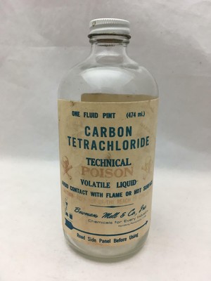
Solvents: Carbon Tetrachloride

Vintage Screw Top Bottle w Label CARBON TETRACHLORIDE Poison Skull ...

Image Source

Carbon tetrachloride is a clear, nonflammable, heavy liquid used as an industrial solvent and reagent with restricted commercial use. It is used in fire extinguishers, dry cleaning agents, grain fumigation, military smokescreens and as a raw material during the manufacturing process of other chemicals (1).


The toxicity profile has long been characterized with increased awareness and subsequent product bans:

  • 1970: banned in consumer products
  • 1978: banned in aerosol products
  • 1986: banned in grain fumigation except for preserving museum artifacts (3)

Today, CCl4 is mainly used in the synthesis of chlorofluorocarbons (CFCs), which are used as heat transfer agents in refrigerating equipment and as aerosol propellants (2).

Exposures

CCl4 vapors can be inhaled occupationally by workers or via direct skin contact while the general population may be exposed by breathing air, smoking cigarettes, consuming food/water and contact with contaminated soil (3).

Mechanism of Action (3, 4)

CCl4 is metabolized primarily in the liver where it is converted to the trichloromethyl radical (CCl3) via the cytochrome p450 isoform CYP2E1. The CCl3 radical then does the following:

  • Lipid peroxidation: free radicals travel to smooth ER and cause lipid peroxidation and other forms of oxidative damage by attacking and destroying polyunsaturated fatty acids. This disrupts the permeability of mitochondria, endoplasmic reticulum and plasma membranes. Without the ability to exchange ions, there is cellular accumulation of calcium resulting in further cell damage. This occurs within the first 10-15 minutes of exposure. 
  • Biotransformation: Converts to trichloromethyl peroxy radical (OOCCl3) and chloroform (CHCl3). Additionally, it is oxidized to produce hexachloroethane (Cl3CCCl3)

These radicals then bind to cellular molecules (nucleic acids, proteins, lipids) and impair processes including cellular respiration, lipid metabolism and fatty degeneration. Free fatty acids then accumulate in the liver resulting in hepatomegaly and other hepatic toxicities. This occurs 10 – 12 hours after exposure.

CCl4 also activates tumor necrosis factor (TNF) alpha, nitric oxide (NO) and transforming growth factors (TGF)-alpha and beta which trigger a signaling cascade resulting in fibrosis and cell death.

Target Organs

Target organs include: endocrine (glands, hormones), liver, CNS, eyes, lungs, liver, kidneys, skin (2).

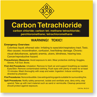
Treatments (2, 3) 

Image Source

  • Anecdote: N-acetylcysteine may reduce severe complications
  • Emergency Treatment:
    • Decontamination
    • Artificial respiration (i.e. demand-valve resuscitator, bag-valve-mask device, or pocket mask)
    • Do not induce vomiting
  • Basic Treatment:
    • Administer oxygen
    • Eye contamination: flush eyes immediately with water or irrigate with 0.9% saline (NS)
    • Ingestion: rinse mouth and administer activated charcoal
    • Dermal Exposure: cover burns with sterile dressings after decontamination
  • Advanced Treatment:
    • Orotracheal or nasotracheal intubation
    • Positive-pressure ventilation techniques
    • Vasopressors if patient is hypotensive
  • Chronic Exposure: no treatment exists

Biomarkers

CCl4 can be detected by gas chromatography in blood, serum, and adipose tissue (3). That being said, any detectable blood level of CCl4 is indicative of exposure. Indirect biomarkers of CCl4 exposure include baseline liver function tests, liver injury tests and renal function tests. In suspected high exposures, clotting studies, chest X rays and electrocardiograms are indicated (3).

CCl4 toxicity Summary

References

(1) Morrison RD, Murphy BL, Doherty RE. 12 – chlorinated solvents. Environmental Forensics. 1964:259-277. http://www.sciencedirect.com/science/article/pii/B9780125077514500343. doi: https://doi.org/10.1016/B978-012507751-4/50034-3.

(2) National Center for Biotechnology Information. PubChem Database. Carbon tetrachloride, CID=5943, https://pubchem.ncbi.nlm.nih.gov/compound/Carbon-tetrachloride#section=Overview (accessed on July 8, 2020)

(3) “Case Study 8: Carbon Tetrachloride Toxicity.” Institute of Medicine. 1995. Environmental Medicine: Integrating a Missing Element into Medical Education. Washington, DC: The National Academies Press. doi: 10.17226/4795.

(4) Weber LW, Boll M, Stampfl A. Hepatotoxicity and mechanism of action of haloalkanes: carbon tetrachloride as a toxicological model. Crit Rev Toxicol. 2003;33(2):105-136. doi:10.1080/713611034

Metals: Lithium

Pictures, stories, and facts about the element Lithium in the ...

Image Source

Lithium (named from “lithos”, Greek word for stone) is a chargeless metal and the simplest xenobiotic. Lithium is a soft, silvery-white alkali metal while also highly reactive and flammable. Lithium is widely distributed on Earth but does not naturally occur in its elemental form due to its high reactivity. Trace amounts of lithium are found in virtually all rocks.

Additionally, trace amounts of lithium have been found in plants, plankton, and invertebrate tissues. Industrial applications include lithium-ion batteries, metal additives, and grease lubricants. Lithium has a limited therapeutic index and has long been used in the treatment of bipolar disease (mania and depression) (1).

Toxicokinetics (1, 2, 4)

  • Bioavailability: rapidly absorbed (~100%)
    • Lithium exerts its effects once it has moved to intracellular compartment
    • Patients with elevated serum levels tend to be asymptomatic
  • Serum Levels: reflect extracellular lithium concentrations
  • Distribution:
    • Widely in total body water and does not bind to serum proteins
    • Tissue distribution follows multiple compartment model
      • Delayed diffusion from the extracellular to the intracellular compartment
  • Absorption/Accumulation:
    • Rapidly taken up by liver, kidney, thyroid, and bone
    • Brain and kidney show highest levels
      • Delayed diffusion in CSF (24 hrs)
      • 80% of lithium that is filtered by kidneys is reabsorbed
    • Overdose: prolonged gastric absorption and clumping from insoluble aggregates may occur (4)
  • Metabolism: Lithium is neither metabolized nor protein-bound.
  • Excretion: 
    • 95% excreted in kidneys
    • ~5% removed via sweat and feces
  • Elimination:
    • Serum elimination half-life can vary 12 to 27 hours
    • Patients with chronic intoxication: half-life up to 48 hours
    • Elderly patients: half-life up to 58 hours
  • Renal Clearance: 
    • Usually 10 to 40 mL/min
    • May be decreased to 15 mL/min
  • Fetal Distribution:
    • Freely crosses placenta and is excreted in breast milk
      • Labeled pregnancy Class D
      • Implicated in increased congenital cardiac defects
      • Reported signs of cyanosis, hypotonia and lethargy in infants of mothers taking lithium

Mechanism of Action (1-3)

The precise mechanism of action of Li+ is currently unknown. An increasing number of scientists have concluded that the excitatory neurotransmitter glutamate is the key factor in understanding how lithium works. Let’s take a look:

Video Source

There are several proposed and confirmed mechanisms of actions for lithium including that noted above. Other mechanisms include:

  • Disrupting glutamate receptors (GluR3) resulting in a decrease (depression) or increase (mania)
  • Non-competitive inhibition of enzyme, inositol monophosphatase (IP) by inhibiting glycogen synthase-3
  • Enhanced deactivation of GSK-3B enzyme resulting in circadian rhythm disruptions and DNA damage
  • Reducing neuronal responsiveness to neurotransmitters due to inhibitory effects of adenylate cyclase and G proteins vital for ion channel opening
  • Stimulating serotonin release from hippocampus

Signs and Symptoms of toxicity (2)

The severity of lithium toxicity is often divided into the following three grades: mild, moderate, and severe.

  • Mild symptoms: nausea, vomiting, lethargy, tremor, and fatigue
  • Moderate intoxication: confusion, agitation, delirium, tachycardia, and hypertonia
  • Severe intoxication: Coma, seizures, hyperthermia, and hypotension

Infographic by Oluwatobi via Canva

Biomarkers/Diagnostic Testing (1)

Once the patient presents to the emergency room, a lithium concentration should be determined, and serial measurements should be performed. Lithium concentration serves as a marker for exposure. 

Infographic by Oluwatobi via Canva

Unique Exposures: Drinking Water (6)

Harari et al., concluded that elevated lithium exposure through drinking water during pregnancy may impair calcium homeostasis, particularly vitamin D. This implication is of a huge public health concern because vitamin D deficiencies have been associated with impaired maternal and fetal health!

In summary, check out this 1-minute video!

Video Source

References:

(1) A. Greller H. Lithium. In: Nelson LS, Howland M, Lewin NA, Smith SW, Goldfrank LR, Hoffman RS. eds. Goldfrank’s Toxicologic Emergencies, 11e. McGraw-Hill; Accessed June 14, 2020. https://accesspharmacy-mhmedical-com.proxy.lib.ohio-state.edu/content.aspx?bookid=2569&sectionid=210274851

(2) Hedya SA, Avula A, Swoboda HD. Lithium Toxicity. [Updated 2019 Nov 13]. In: StatPearls [Internet]. Treasure Island (FL): StatPearls Publishing; 2020 Jan-. Available from: https://www.ncbi.nlm.nih.gov/books/NBK499992/

(3) Lithium cation. https://www.drugbank.ca/drugs/DB01356. Updated 2007. Accessed June 14, 2020.

(4) Decker BS, Goldfarb DS, Dargan PI, et al. Extracorporeal Treatment for Lithium Poisoning: Systematic Review and Recommendations from the EXTRIP Workgroup. Clin J Am Soc Nephrol. 2015;10(5):875‐887. doi:10.2215/CJN.10021014

(5) Pérez-Granados, Ana & Vaquero, MP. (2002). Silicon, aluminium, arsenic and lithium: Essentiality and human health implications. The journal of nutrition, health & aging. 6. 154-62.

(6) Harari F, Åkesson A, Casimiro E, Lu Y, Vahter M. Exposure to lithium through drinking water and calcium homeostasis during pregnancy: A longitudinal study. Environmental Research. 2016;147:1-7. http://www.sciencedirect.com/science/article/pii/S0013935116300305. doi: https://doi.org/10.1016/j.envres.2016.01.031.

Pesticides: Rotenoids

What is Rotenone?

Image Source

Rotenone is a naturally occurring compound found in the roots of several plant species of the pea (Leguminosae) family. Being the first member of the rotenoid family, its use includes:

  • Insecticide (insect control) in home gardening
  • Piscicide (fish eradication) for freshwater management
  • Acaricide (lice, scabies, mites and tick control) for pets

It is generally classified as a botanical insecticide and it is a commonly used pesticide.

Rotenone is used alone or in combination with pyrethrin, pyrethrum, and piperonyl butoxide to control a variety of insects on food crops (5). Rotenone is a moderately hazardous Class II pesticide. In the U.S. and Canada, rotenone is mainly used as a piscicide.

Biotransformation

Please see the metabolic scheme of the major biotransformation pathways of rotenone by hepatic microsomes

Image Source

  • Metabolites: hydroxyrotenone, rotenolone I and II, dihydroxyrotenone
  • Physicochemical Properties:
    • High melting point
    • Insoluble in water
    • Soluble in organic solvents
    • Highly lipophilic
  • Stability:
    • Decomposes rapidly when exposed to light and air
      • Loses toxicity within days
  • Formulations:
    • Dust – to control beetles and aphids on produce
    • Wettable powder – to control parasitic mites, lice, and ticks
    • Emulsion – used for undesirable fish in water management
    • Formulated with other pesticides for wide-ranging effects

Infographic by Oluwatobi Clement via Canva

Infographic by Oluwatobi Clement via Canva

Mechanism of Action

Remember that electron transfer is needed in order to pump protons (H+ ions) across the gradient from the mitochondrial matrix into the cell. The resulting protocol gradient triggers the molecular rotation of the ATP synthase, producing ATP. Need a refresher? Let’s revisit the electron transport chain (ETC):

(Video Source)

Rotenone interferes with the electron transport chain by inhibiting the transfer of electrons from iron-sulfur centers in Complex I to ubiquinone. This inhibition interferes with NADH during ATP synthesis. Complex I is then unable to pass off its electron to Coenzyme Q (CoQ), creating a back-up of electrons within the mitochondrial matrix. In mammals and fish, rotenone inhibits the oxidation of NAD and substrates glutamate, alpha-ketoglutarate and pyruvate.

Image source

In addition to its effects on the ETC, rotenone interferes with reactive oxygen species (ROS) by inducing apoptosis. Rotenone inhibits the ATP production and increased generation of ROS causing the inhibition of neuronal activity (2). The increased ROS results in increased calcium and activation of calcium-sensitive potassium channels. This is called nitrosative stress.

Image Source

Other actions of rotenone include:

  • Increased nitric oxide and malondialdehyde levels
  • Global cellular impairment
  • Aggregation of alpha-synuclein and polyubiquitin
  • Activation of astrocytes and microglial cells
  • Neuroinflammatory reaction
  • Glutamate excitotoxicity
  • Oxidative stress
  • Microtubule assembly and spindle formation in mitosis (4) resulting in chromosomal aberrations

Which organs does it target?

  • Cutaneous membrane (skin)
  • Mucous membranes:
    • Respiratory tract (inhalation)
    • Digestive tract (digestion)
    • Eye irritant (eye contact)
  • Central Nervous System
  • Peripheral Nervous System: causes anesthetic effect when in contact with nerve axons
  • Skeletal and Smooth muscle

Signs and symptoms of toxicity

Parkinson’s Disease

In addition to the effects noted earlier, the following activities have been observed when rotenone is administered experimentally:

  • Degeneration of striatal-nigral dopaminergic neurons similar to neurotoxin MPP (a well-known Parkinsonism-causing chemical and Complex I inhibitor)
  • Degeneration of dopaminergic neurons in rotenone-evoked Parkinsonism
  • Production of protein inclusions, similar to Lewy bodies
    • Stain positive for ubiquitin and alpha-synuclein

Ultimately, no strong evidence of Parkinson disease-like signs or neurodegenerative pathologies have been found with rotenone exposure. However, a strong association between rotenone and increased risk of PD has been noted.

Click (1)(2) or (3) for more information.

NIH News Release (here)

Biomarkers

Rotenone is 100x more toxic when administered IV than oral due to poor absorption from GI tract.

Biomarkers of rotenone exposure include:

  • Residue detection of rotenone and/or its metabolites in blood, urine, feces, or liver
    • Determined using HPLC with a fluorescence detector or Liquid Chromatograph-Mass Spectrometer (LC-MS-MS)
  • Metabolites (in decreasing order of toxicity):
    • Hydroxyrotenone: have reduced inhibitory activity in insect and rat liver mitochondria
    • Rotenolone I and II
    • Dihydroxyrotenone
  • Characteristic toxicological symptoms
  • Histopathological changes

Infographic by Oluwatobi Clement via Canva Source

Let’s Test Your Knowledge!

  1. Which of the following metabolites causes the most systemic toxicities?
    1. Hydroxyrotenone
    2. Rotenolone I
    3. Dihydroxyrotenone
    4. Rotenolone II
  2. Where is rotenone mainly metabolized?
    1. Dermally (Sweat)
    2. Feces
    3. Exhalation
    4. Urine
  3. Which of the following vitamins is an experimental therapy for acute rotenoid exposure?
    1. Vitamin D
    2. Vitamin B12
    3. Vitamin E
    4. Vitamin K
  4. Which of the following techniques are used to quantitatively detect rotenone biomarkers in the blood?
    1. GC and MS
    2. FPLC and ELISA
    3. HPLC
    4. HPLC and LC-MS-MS
  5. For an accidental ingestion, one should induce vomiting within 1 hour of exposure.
    1. True
    2. False
  6. Which of the following is not a rotenoid metabolite?
    1. Dihydroxyrotenone
    2. Trimethylrotenone II
    3. Rotenolone I
    4. Hydroxyrotenone
  7. Which of the following cardiac toxicities are observed in chronic exposures?
    1. Arrythmia
    2. Prolonged QT interval
    3. Tachycardia
    4. None of the Above

Answers:

  1. A
  2. B
  3. D
  4. D
  5. B
  6. C
  7. D

Resources

  1. Costa LG. Toxic Effects of Pesticides. In: Klaassen CD. eds. Casarett and Doull’s Toxicology: The Basic Science of Poisons, Eighth Edition New York, NY: McGraw-Hill; 2013. http://accesspharmacy.mhmedical.com.proxy.lib.ohio-state.edu/content.aspx?bookid=958&sectionid=53483747. Accessed May 25, 2020.
  2. Yee AG, Freestone PS, Bai J, Lipski J. Paradoxical lower sensitivity of locus coeruleus than substantia nigra pars compacta neurons to acute actions of rotenone. Experimental Neurology. 2017;287:34-43. http://www.sciencedirect.com.proxy.lib.ohio-state.edu/science/article/pii/S0014488616303338. doi: https://doi-org.proxy.lib.ohio-state.edu/10.1016/j.expneurol.2016.10.010.
  3. Newhouse K, Shih-Ling Hsuan, Chang SH, Beibei Cai, Yupeng Wang, Zhengui Xia. Rotenone-Induced Apoptosis Is Mediated By p38 And JNK MAP Kinases In Human Dopaminergic SH-SY5Y Cells. Toxicological Sciences. 2004;79(1):137-146. doi:10.1093/toxsci/kfh089.
  4. Krieger, R. (ed.). Handbook of Pesticide Toxicology. Volume 2, 2nd ed. 2001. Academic Press, San Diego, California., p. 1186
  5. Bavli D, Prill S., Ezra E, Levy G., Cohen M.,Vinken M.,Vanfleteren J., Jaeger M., Nahmias Y.,
  6. National Center for Biotechnology Information. PubChem Database. Rotenone, CID=6758, https://pubchem.ncbi.nlm.nih.gov/compound/Rotenone (accessed on May 27, 2020)
  7. Gosalvez, M. (1983). Carcinogenesis with the insecticide rotenone. Life Sci. 12, 809-816
  8. Greenman, D., Allaben, W., Burger, G., Kodell, R. Bioassay for carcinogenicity of rotenone in female wistar rats. Fundamental and Applied Toxicology. 1993;20:383-390.