NEW OPENING: March 2024
Postdoc Position available – High Resolution Retinal Imaging
Postdoctoral Scholar in High Resolution Retinal Imaging
A NIH funded Post Doctoral Scholar position is available in the laboratory of Dr. Nathan Doble in the College of Optometry/Department of Ophthalmology and Visual Science at The Ohio State University (OSU). The successful candidate will work within a highly collaborative research environment focused on the development and use of novel high-resolution optical instrumentation for the study of retinal structure and function. The candidate will be expected to assist with construction and validation of novel instrumentation as well as the collection and analysis of the experimental data; preparation of research results for publication and talks; writing the scientific manuscripts, progress reports, and participation in grant writing.
This particular project will be conducted in collaboration with the University of California, Davis to design and implement full-field optical coherence tomography (FF-OCT) for the study of structure and function of individual retinal neurons in the living human and murine eyes. As part of this work we have recently demonstrated a parallel line scanning ophthalmoscope capable of imaging the eye at 500 Hz provided exceptional imaging of single red blood cells (Lee et al. Biomedical Optics Express, 2024).
Experience:
Candidate should have experience in the field of biomedical imaging or optics. Specifically, a background in development or application of optical instrumentation for the human retinal imaging would be particularly advantageous. Additionally, a high degree of proficiency in programming (e.g. Matlab, LabView, Python, C++, GPU or equivalent) and hardware control (e.g. high speed cameras and digital mirror devices) is required. Excellent communication and writing skills with a good balance of independence and collegiality in research are essential.
Education:
Doctorate in physics, biomedical physics, biomedical engineering, or electrical engineering.
Interested candidates should submit a cover letter, resume, a statement of research skills and interests, and contact details of three referees (please describe relationship to each). Applicants should apply at:
https://osu.wd1.myworkdayjobs.com/OSUCareers/job/Columbus-Campus/Post-Doctoral-Scholar_R100061-1
Job number is R100061.
The College offers a competitive salary and excellent benefits.
Imaging Individual Red Blood Cells In-Vivo
A new high speed line scanning partially confocal imaging system operating at 500 Hz allows for excellent visualization of the movement of red blood cells in the living human retina. Please see our recent publication by Lee et al. in Biomedical Optics Express 2024.
Imaging Retinal Ganglion Cells (RGCs) In-Vivo
We are one of a few AO groups with the ability to image individual neurons within the inner retina. Figure 1 below shows a scan through video of a normal control moving posterior from the nerve fiber layer. We developed registration code that registers 60-80 AO-OCT volumes in order to have sufficient contrast to see the individual RGC soma. Contrast has been optimized for the RGC layer.
Figure 1 – In-vivo images of RGC soma using our AO-OCT-SLO
Figure 2 shows AO-OCT B-scans and en-face images from a primary open angle glaucoma subject at day 1 and 4 months later. Note the black lesions in the inner nuclear layer. Please see our recent Journal of Glaucoma paper for more information.

Figure 2 – AO-OCT B-scans and en-face images from a POAG subject
Age-Related Macular Degeneration (AMD)
Another major project is imaging patients with AMD to determine the photoreceptor health over drusen. Figure 3 shows a montaged AO-OCT image (linear display) for an early stage AMD subject from 6 degree superior retina to 1 degree inferior retina. Cone photoreceptors are intact over the smaller drusen but the outer segment is absent over the larger deposits. Scale bar is 100 µm in both directions.

Figure 3 – Montaged AO-OCT image (linear display) for an early stage AMD subject from 6 degree superior retina to 1 degree inferior retina. Cone photoreceptors are intact over the smaller drusen but the outer segment is absent over the larger deposits. frames. Scale bar is 100 µm in both directions.
Imaging Rod Photoreceptors
Our group was also the first the demonstrate the ability to image the rods in-vivo (ARVO 2004, Optics Letters 2011). More recently we have sought to extent this capability to the mid-periphery and this work as published in Eye (2016). Figure 4 shows images from our AO-SLO. The ability is particularly important in the study of AMD and retinitis pigmentosa (RP) as the fisrt signs are usually in the periphery.
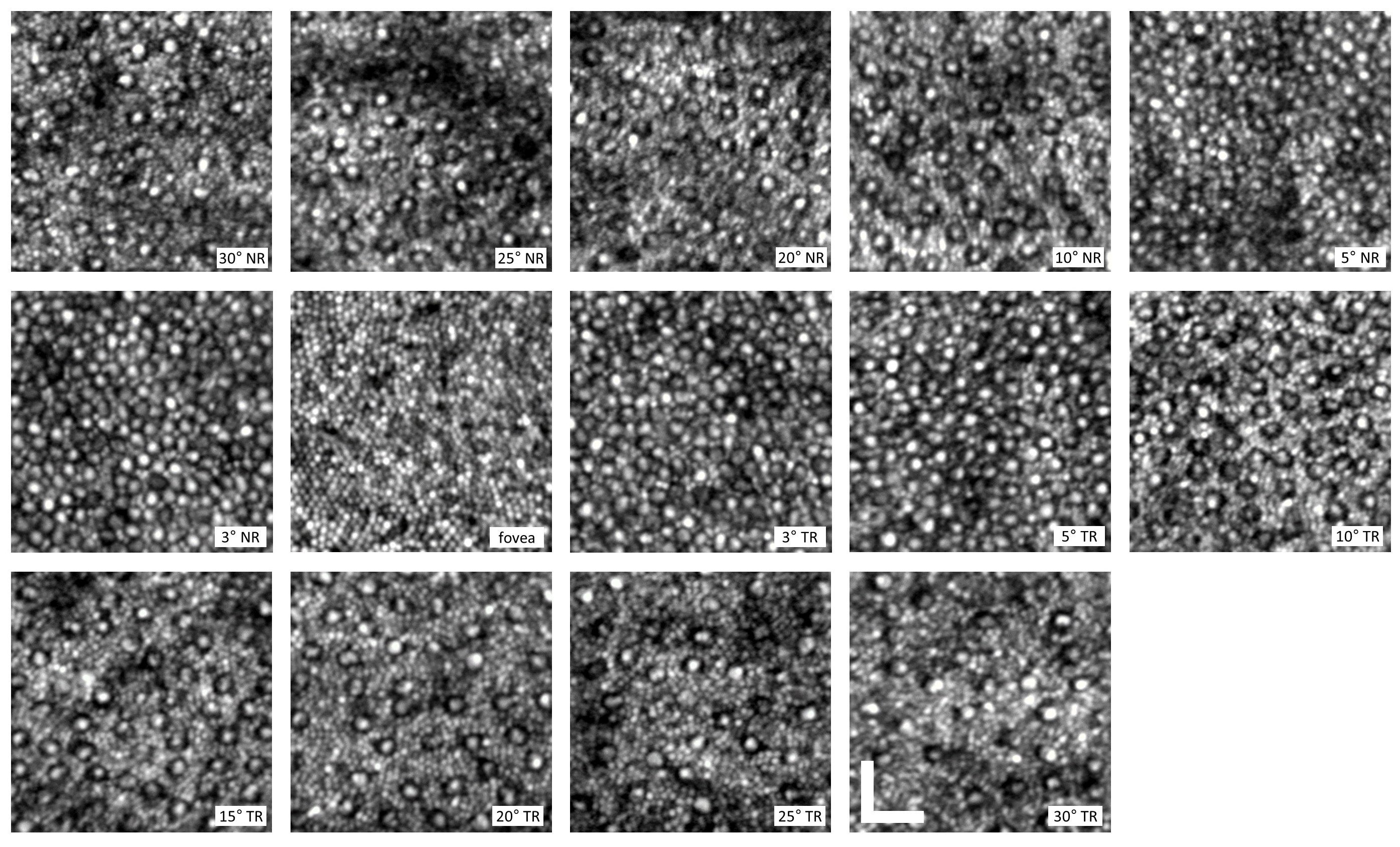
Figure 4. In vivo human rod and cone AO-SLO images from 30 degrees in the nasal retina to 30 degrees in the temporal retina. The cones are the larger structures in between the smaller rods. The variation in density with retinal eccentricity is clearly evident. Scale bar is 25 microns. Published in the journal Eye 2016.