
What Is It? | Facts in Depth | For the Professional Diagnostician
Cucurbit Diseases | Cucurbit Downy Mildew
Cucurbit Downy Mildew
Symptoms
Leaves:
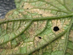
Chlorotic, yellow to olive lesions, often vein-delimited, are scattered across the leaf. Lesions may have a greasy sheen. As disease progresses, lesions become tan-brown and necrotic. On watermelon, lesions are more circular, and on melon, lesions are not always vein-delimited.


Signs
Fuzzy, white to dark gray-purple sporulation can be seen on the undersides of lesions. Dichotomously branching sporangiophores bear oval, papillate sporangia. Sporangia can be hyaline to gray-purple depending on age. Sporangia are 20-40 μm long and 14-25 μm wide. Oospores (22-42 μm in diameter) can be produced by P. cubensis, but are rarely seen.


Often Confused With
- Angular leaf spot: Check for bacterial streaming. No streaming will be present if it is downy mildew.
- Cercospora leaf spot: Place samples in a moisture chamber and look at what type of sporulation is present microscopically.
- Powdery Mildew: These diseases can co-occur and chlorosis from powdery mildew can be confused with downy mildew. Check sporulation under the microscope.
Isolation Media
P. cubensis is an obligate pathogen and cannot be cultured. If pathotype needs to be determined, the pathogen can be propagated on immature, susceptible plants and tested on differential lines. Six pathotypes have been described. For more information on these assays, see: Lebeda, A. and Widrlechner, M.P. 2003. A set of Cucurbitaceae taxa for differentiation of Pseudoperonospora cubensis pathotypes. Journal of Plant Disease Protection 110: 337–349.
Rapid Diagnostic Tests
The distinctive sporangiophores of P. cubensis make diagnosis relatively simple and make molecular tests unnecessary for diagnosis. If sequencing is necessary, semi-specific primers for the ITS region of Peronosporales and Pythiales can be used.
DC6 5’GAGGGACTTTTGGGTAATCA 3′
ITS4 5′ TCCTCCGCTTATTGATATGC 3