Gila Monster Toxicology

Gila Monster Overview

Gila Monster | National Geographic

Image 1 (Source

The Gila Monster is a large, venomous, desert-dwelling lizard that, in the United States, is generally found in the arid regions of Arizona, Utah, New Mexico, southern California, and southern Nevada¹. These rocky-terrain-dwelling lizards have a diet primarily consisting of smaller birds as well as eggs of various organisms¹. Unlike other venomous creatures that utilize hollow fangs, the gila monster employs capillary action with grooved teeth to bite venom into its prey. Due to this more primitive mechanism of delivery, the gila monster is known to latch on to prey and continue to chew, thus helping to force sufficient venom into the prey through said capillary action. To the general population, even seeing a gila monster is extremely rare, thus making human envenomations highly unlikely. The following video provides an interesting look at the gila monster skull, why human envenomations are so rare, and the process of milking the venom for research.

https://youtu.be/tajSlpFZRaY?t=137

Video 1 (Source)

As with most other venomous animals, gila monster venom contains a number of components. Important toxicologic components include serotonin, amine oxidase, phospholipase A, substances promoting the release of bradykinin, helodermin, hyaluronidase, and exendin 4. As will be discussed later, exendin 4 has been revolutionary and crucial to the creation of a drug class for type-2 diabetes treatment known as GLP-1 analogues.

Mechanisms of Toxicity

Image 2 (Source)

The mechanism of toxicity for each aforementioned venom component will be described individually below. As venoms are complex mixtures of many chemicals, exact effects are often unclear. Overall symptoms of gila monster envenomation will be discussed later.

Serotonin

Serotonin in venom exhibits numerous physiologic consequences, including potent vasodilation, increased platelet aggregation, and pain production through direct irritation of tissues. Serotonin predominantly produces these effects by direct agonism of the various serotonin receptors found throughout the body. Vasodilation is mediated via agonism of endothelial 5-HT receptors in the blood vessels by serotonin, causing an increased release of endothelial-derived relaxing factor leading to the release of nitric oxide, a potent vasodilator. Serotonin also promotes the release of prostaglandin I2, a known vasodilator. Other effects, such as that of pain production, still require further elucidation.

Amine Oxidase

Amine oxidases are thought to cause damage due to their enzymatic, oxidative functions. This oxidation of proteins may create reactive and toxic oxygen species, such as hydrogen peroxide, leading to direct cell toxicity and apoptosis²,³.

Phospholipase A

Phospholipase A serves to regulate fatty acid metabolism in the cell, cleaving fatty acid tails from glycerol molecules. Cleavage of these fatty acids releases arachidonic acid, leading to marked inflammation and tissue damage².

Bradykinin

Bradykinin is crucial in the inflammatory pathway, inducing the production of various prostaglandins that cause marked vasodilation, increased vascular permeability, swelling, and tissue irritation.

Helodermin

Helodermin, a non-enzymatic peptide, is thought to produce hypotension via the activation of certain potassium channels in the endothelium². However, the effects of helodermin are still not fully described.

Hyaluronidase

This ubiquitous venom component is well-known for its effects in breaking down hyaluronic acid derivatives, increasing membrane permeability and allowing for greater venom penetration into tissues².

Exendin 4

Exendin 4 is a notable gilatoxin due to its similarities to endogenous human GLP-1. Via a glucose-dependent process, GLP-1 increases insulin secretion and suppresses glucagon secretion, leading to lowered blood sugars. However, as this process is glucose-dependent, hypoglycemia is not commonly associated with GLP-1. This peptide also exerts effects on appetite as well as gastric emptying. GLP-1 produces a direct suppression of POMC neurons in the appetite control center of the brain, leading to suppression of appetite. GLP-1 also antagonizes gut motility, delaying gastric emptying and promoting satiety for a longer period of time (see reference 4 for more detail). Overall, this fraction of venom may be heavily implicated in the nausea and vomiting produced by envenomation². Below is a figure from the American Diabetes Association depicting the various actions of GLP-1.

Image 3 (Source)

Toxicokinetics

As discussed above, the predominant route of exposure to gila monster venom is envenomation via bite wounds. This route bypasses other common toxin exposure sites, including the lungs and GI tract. As such, discussion of absorption is less applicable. The various components of gila monster venom are metabolized and excreted via different means. Serotonin is primarily removed from the plasma via reuptake into cells. While either outside the cell or after reuptake, serotonin is degraded by monoamine oxidase (MAO), an enzyme that cleaves the left amino chain. This metabolite, known as 5-HIAA is excreted via the urine once it has undergone phase-2 glucaronidation or sulfation (see reference 5 for more detail). Bradykinin may be metabolized by a number of enzymes, namely angiotensin-converting enzyme (ACE), to inactive metabolites. One such metabolite, kallirenin, is excreted via the urine. Little pharmacokinetic data is available on other components of gila monster venom, including helodermin, hyaluronidase, amine oxidase, and phospholipase A. Exendin-4, the GLP-1 analogue, is better studied, with an estimated half-life of 18-41 minutes for IV administration in rat models (see reference 6). This is slightly longer than the half-life of endogenous GLP-1, which has a half life of about 2 minutes, primarily via degradation by endogenous DPP-4. While little data is available on Exendin-4, the GLP-1 agonist most similar in structure to it, Exenatide (Byetta), undergoes minimal enzymatic metabolism. Exenatide is instead excreted predominantly in the urine (source 7). Overall, the toxicokinetics of various components of gila monster venom require further study and elucidation.

Genetic Variations

No information on the effects of genetic variations on gila monster toxicology could be found.

Carcinogenicity

No information on the carcinogenic effects of gila monster venom could be found.

Biomarkers

Unfortunately, there are currently no reliable biomarkers to assess a patient’s exposure to gila monster venom. Currently, diagnosis is made via oral patient history.  As mentioned before, gila monster envenomation of humans is extremely rare.

Symptoms of Toxicity

Low Blood Pressure Stock Illustrations – 809 Low Blood Pressure Stock Illustrations, Vectors & Clipart - Dreamstime

Image 4 (Source)

The first symptom of gila monster envenomation is localized pain and swelling due to the various tissue-damaging toxins of the venom (bradykinin, serotonin, etc.). Other symptoms then appear, including nausea, vomiting, weakness, sweating, and hypotension². If untreated, the bite wound may also become infected, resulting in skin and soft tissue infections or more serious complications such as bacteremia.

Managing of Poisoned Patients

Currently, no antivenom is available to treat gila monster envenomation. Treatment is primarily supportive, including fluid resuscitation, pressors (dopamine, midodrine, etc.), pain management, wound cleaning, and infection prophylaxis/treatment².

Historical Relevance 
Byetta, Bydureon & Bydureon BCose | Usage & Dosage

Image 5 (Source)

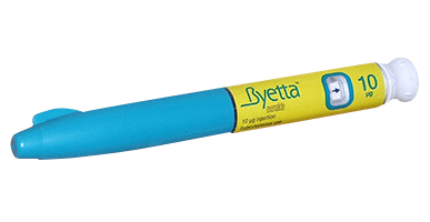
As mentioned before, the most historically-relevant link to gila monster venom comes in the GLP-1 drug class for treatment of type-2 diabetes mellitus as well as obesity. The first drug in this class based on Exendin-4 isolated from the gila monster, Byetta (shown above), was approved by the FDA in 2005. since then, numerous other GLP-1 analogues have come to market, including Ozempic, Victoza, Bydureon, and even Rybelsus, an orally-active form of semaglutide (Ozempic). While the half-life of endogenous GLP-1 is only a few minutes, these drugs have various modifications to decrease metabolism and slow release into the blood stream. This has allowed administration to change from twice daily with Byetta to once weekly for Ozempic. These medications have revolutionized type-2 diabetes care, where an impaired GLP-1 response to meals has been noted. Due to their lack of association with hypoglycemia, positive effects on satiety and weight loss, and infrequency of administration, these GLP-1 analogues are now the first injectable medication of choice indicated by ADA guidelines for type-2 diabetics. For more information on this drug class, please visit the ADA’s Standards of Medical Care in Diabetes – 2020 (source 7).

References

  1. Gila monster. Smithsonian’s National Zoo. https://nationalzoo.si.edu/animals/gila-monster. Published June 29, 2018. Accessed July 28, 2021.
  2. Watkins JB, III. Toxic Effects of Plants and Animals. In: Klaassen CD. eds. Casarett and Doull’s Toxicology: The Basic Science of Poisons, Eighth Edition. McGraw Hill; Accessed July 27, 2021. https://accesspharmacy-mhmedical-com.proxy.lib.ohio-state.edu/content.aspx?bookid=958&sectionid=53483751
  3. Bhattacharjee P., Mitra J., Bhattacharyya D. (2017) L-Amino Acid Oxidase from Venoms. In: Gopalakrishnakone P., Cruz L., Luo S. (eds) Toxins and Drug Discovery. Toxinology. Springer, Dordrecht. https://doi.org/10.1007/978-94-007-6452-1_11
  4. https://spectrum.diabetesjournals.org/content/30/3/202
  5. S. Athar, Serotonin and Tryptophan, Editor(s): Katie Kompoliti, Leo Verhagen, Metman, Encyclopedia of Movement Disorders, Academic Press, 2010, Pages 104-108, ISBN 9780123741059, https://doi.org/10.1016/B978-0-12-374105-9.00001-0.
  6. Parkes, D., Jodka, C., Smith, P., Nayak, S., Rinehart, L., Gingerich, R., Chen, K. and Young, A. (2001), Pharmacokinetic actions of exendin-4 in the rat: Comparison with glucagon-like peptide-1. Drug Dev. Res., 53: 260-267. https://doi.org/10.1002/ddr.1195
  7. American Diabetes Association (ADA). Standards of medical care in diabetes–2020. Diabetes Care. 2020;43(suppl 1):S1-S212. https://care.diabetesjournals.org/content/43/Supplement_1. Accessed January 22, 2020.

Benzene Toxicology

Benzene Overview

Image 1 (Source)

Benzene is a highly ubiquitous, volatile, flammable solvent produced from petroleum with extensive industrial uses. Apart from its use as a solvent, benzene is used to produce other materials including styrofoam plastics, cumene resins, glues, paints, and nylon fiber. Naturally, benzene can be found in crude oils and combustion products, such as volcanic gasses, forest fire smoke, and cigarette smoke. Benzene exposures occur in the general population daily, mainly through inhalation. Of particular interest is that half of all benzene exposures come directly from smoking or inhaling cigarette smoke. As benzene is can be released by automobiles, manufacturing, and various products containing it, exposures are generally greater in cities than in more rural areas. Oral consumption of benzene is a far smaller source of exposure than inhalation. If a water source is contaminated, significant dermal exposure to benzene can occur when bathing or showering (although this is not the main route of exposure). Overall, those at greatest risk of exposure to higher levels of benzene are those working in industries that manufacture or use benzene products. Benzene concentrations in the environment are as follows:

  • Outdoor Air = 0.02 – 34 parts per billion (heavily dependent on location)
    • Average = 1.9 parts per billion
  • Indoor Air, Home (Non-smoker) = 2.2 parts per billion
  • Indoor Air, Home (Smoker) = 3.3 parts per billion
  • Indoor Air, Bar (Smoker) = 8.08 – 11.3 parts per billion

All values reported per the EPA (Source)

The table below is included in the U.S. Department of Health and Human Services Agency for Toxic Substances and Disease Registry (ATSDR) profile for benzene. Major U.S. cities and the average air concentrations of benzene are listed.

Image 2 (Source)

In addition to benzene that is produced upon combustion, tobacco products contain many other detrimental chemicals. See this video by the FDA for more information:

https://www.youtube.com/watch?v=EXdxl0yH904

Video 1 (Source)


Mechanisms of Toxicity

Image 3 (Source)

While two most concerning effects associated with systemic benzene toxicity are hematotoxicity and leukemogenicity, benzene is also a known direct irritant, causing skin, eye, and lung tissue irritation and damage. Benzene exposures have also been linked to female reproductive abnormalities, acute GI distress following high oral exposure, and acute pulmonary exposure to high atmospheric levels of benzene. To date, only the hematotoxic and leukemogenic effects have been studied sufficiently to propose a mechanism of toxicity.

Hematotoxicity and Leukemogenicity

According to mechanistic studies in mice, benzene’s hepatic metabolites readily distribute into the bone marrow through passive diffusion due to their small size and lipophilicity. Of particular concern are the phenolic metabolites of benzene (discussed in more detail below), which possess the capability to induce oxidative stress, directly damaging proteins and DNA of stem/progenitor cells. This stem/progenitor cell damage can cause apoptosis and impedes the formation of new red and white blood cells. DNA damage induced by benzene’s metabolites is also extensively linked to leukemia (cancer of the blood-forming organs). Overall, benzene’s more potent metabolites create oxidative stress, inducing DNA damage and causing different manifestations of hematic, lymphatic, and leukemogenic dysfunction.

Carcinogenicity

As described above, benzene is a known human carcinogen and, as such, is classified as Group 1 by the IARC. Benzene exposures have been linked to hematopoietic cancers, lymphatic cancers, and various leukemias. Benzene is carcinogenic through all routes of exposure, though inhalation is still the most prevalent of these.

The following is a video with more information detailing causes of leukemia, with benzene exposure and smoking featured extensively.

Video 2 (Source)

Toxicokinetics

Benzene is readily absorbed via all routes of exposure due to its low molecular weight (78.11 g/mol), small size, and relative lipophilicity (logP = 2.13). Due to these properties, benzene is postulated to cross membranes via passive diffusion and be readily bound to plasma proteins. Benzene is widely distributed into fatty tissues due to its lipophilicity. As discussed above in mechanisms of toxicity, benzene’s hepatic metabolites are heavily implicated in its toxic effects. Benzene is rapidly metabolized in the liver via cytochrome p450 enzymes, namely CYP2E1, to phenolic compounds such as phenol, catechol, hydroquinone, 1,2,4-benzenetriol, and 1,2- and 1,4-benzoquinone as well as benzene oxide. Of these, the phenolic compounds are most notable for pathogenesis of the diseases caused by benzene exposure and are found in the bone marrow after inhalation exposures. Below is a figure from Cancer Epidemiology Biomarkers and Prevention detailing many of these toxic metabolites of benzene.

Image 4 (Source)

Excretion

Inhaled, unmetabolized benzene is predominantly excreted via exhalation in a triphasic manner. Percentage of benzene exhaled unchanged appears highly dependent on dose in both rat and human models. In a study with 23 volunteers exposed to 47-110 ppm benzene-containing air for multiple hours, the percent of the benzene dose exhaled ranged from 16.4 to 41.6 percent. Rat and mouse studies also indicate that the higher the dose inhaled, the greater percentage of the benzene dose is exhaled. Phenolic benzene derivatives are primarily conjugated to glucaronides and sulfates and subsequently excreted via the urine in a biphasic manner. A very small amount (<3.5% for rats and <9% for mice) of benzene and its derivatives is excreted fecally. Benzene’s tissue half life is variable due to its multi-phasic elimination, with a range of 0.4 – 1.6 hours. Conjugated phenols have a half life of around 1 hour. In high-lipid tissues in some studies, benzene’s half life has been proposed to be around 24 hours. Overall, more studies are necessary to determine accurate half-life values for benzene in humans.

Genetic Variations

As described above, CYP450 metabolism of benzene to phenolic and other derivatives is incredibly important for its toxic effects. Following this reasoning, mice were pre-treated with CYP450 inhibitors before inhalation exposure to benzene. This inhibition of the ‘activating’ enzymes led to decreased genotoxicity, a hallmark for benzene. Therefore, it stands to reason that those with CYP450 deficiencies, especially CYP2E1, may have decreased benzene toxicity due to chronic inhalation exposure. More tests are necessary to fully elucidate the effects of genetic variations on benzene toxicity.

Biomarkers

If inhaled, unmetabolized benzene can be detected in expired air as well as, to some extent, in the urine. As benzene is primarily eliminated via urinary excretion of toxic metabolite conjugates, urinary phenol,trans,trans-muconic acid, and S-phenylmercapturic acid have all seen use to assess occupational exposures. However, these biomarkers appear to be useful in cases of prolonged occupational exposure or acute exposure to high levels of benzene. The accuracy of these markers to assess environmental exposures is less clear.


Symptoms of Toxicity

Image 5 (Source)

Symptoms of benzene toxicity are dependent on the route and severity of exposure. Below are symptoms of toxicity broken up by these methods.

Acute Inhalation Exposure

Acute exposure to levels far higher than are found in the environment (10,000 to 20,000 parts per million, normal exposure values around 0.0019 parts per million) results in neurologic symptoms. Symptoms include drowsiness, confusion, dizziness, lightheadedness, headaches, tachycardia, and tremors. Symptoms typically resolve upon exposure to fresh, uncontaminated air.

Acute Oral Ingestion

Symptoms of GI distress after exposure to high levels or oral benzene are most likely related to its direct irritant effects. GI distress may be accompanied by unexplained neurologic symptoms to include: nausea, vomiting, dizziness, drowsiness, convulsions, and tachycardia.

Acute Dermal/Optical Exposure

Direct skin contact with benzene may cause symptoms of redness, sores, and pain. Benzene exposure in the eyes causes direct irritation, pain, inflammation, and corneal damage.

Chronic Inhalation Exposure

As discussed extensively above, benzene is a known carcinogen that may cause hematopoietic cancers. Mutations in the proteins and DNA of stem/progenitor cells may result in reduced numbers of lymphatic cells, decreased red blood cells (anemia), fatigue, weakness, dizziness, lightheadedness, shortness of breath, or excessive bleeding. Symptoms of anemia are depicted in Image 6 below. Chronic inhalation exposure has also been associated with reproductive abnormalities in women. Symptoms may include menstrual irregularities and decreased ovary size.

Image 6 (Source)

Management of Poisoned Patients

The treatment of acute exposures to benzene focuses on exposure control. If exposed dermally or optically, the affected area should be rinsed (with sterile water or sterile normal saline) and any contaminated clothing removed. For high-level inhalation exposure, emphasis should be made on moving the patient to an environment with fresh, uncontaminated air. Symptoms of these type of exposures generally improve with source control. Should respiratory distress occur after an inhalation exposure, 100% humidified oxygen and mechanical ventilation may also be used. Once distributed, there are currently no methods to reduce total body burden of benzene. Myelotoxic effects of benzene exposure has been mediated using the non-steroidal anti-inflammatory (NSAID) drug indomethacin in mice. Developing research indicates that TNF may be useful in interfering with hematotoxic effects of benzene. However, far more research is required to optimize treatment of benzene toxicity and prevent the hematotoxic and leukemogenic effects.

Historical Exposure

One historical exposure of note stands out in the literature. Studies were performed on workers exposed to benzene in three separate Pliofilm factories in Ohio. In the 1987 study report, 1,165 white male employees were studied for their occupational exposure to benzene. Analyses of the data from these studies showed an undeniable correlation between benzene exposure and mortality related to leukemia. This study continued for another 15 years, concluding that the risk of these adverse effects dropped as the time since last exposure increased. Similar studies in China have also solidified the leukemogenic potential of benzene.


References

  1. Agency for Toxic Substances and Disease Registry (ATSDR). 2008. Toxicological profile for Benzene. Atlanta, GA: U.S. Department of Health and Human Services, Public Health Service.
  2. Yoon BI, Hirabayashi Y, Kawasaki Y, Kodama Y, Kaneko T, Kim DY, Inoue T. Mechanism of action of benzene toxicity: cell cycle suppression in hemopoietic progenitor cells (CFU-GM). Exp Hematol. 2001 Mar;29(3):278-85. doi: 10.1016/s0301-472x(00)00671-8. PMID: 11274754.
  3. McHale CM, Zhang L, Smith MT. Current understanding of the mechanism of benzene-induced leukemia in humans: implications for risk assessment. Carcinogenesis. 2012;33(2):240-252. doi:10.1093/carcin/bgr297.
  4. National Center for Biotechnology Information. PubChem Compound Summary for CID 241, Benzene. https://pubchem.ncbi.nlm.nih.gov/compound/Benzene. Accessed July 5, 2021.
  5. Kim S, Vermeulen R, Waidyanatha S, et al. Modeling Human Metabolism of Benzene Following Occupational and Environmental Exposures. Cancer Epidemiology Biomarkers & Prevention. 2006;15(11):2246-2252. doi:10.1158/1055-9965.epi-06-0262.
  6. Iron-refractory iron deficiency anemia: MedlinePlus Genetics. MedlinePlus. https://medlineplus.gov/genetics/condition/iron-refractory-iron-deficiency-anemia/#causes. Published August 18, 2020. Accessed July 5, 2021.
  7. U.S. Food and Drug Administration. Chemicals in Every Puff of Cigarette Smoke – Combustion Stage [Video]. YouTube. https://www.youtube.com/watch?v=EXdxl0yH904. Published February 13, 2017. Accessed July 5, 2021.
  8. UC Berkeley Events speaker Martin T. Smith. Finding the causes of leukemia [Video]. YouTube. https://www.youtube.com/watch?v=07_Snolni-g. Published June 21, 2012. Accessed July 5, 2021.


Aluminum Toxicology

Aluminum Overview

Aluminum.png

Image 1 (Source)

Aluminum is one of the most abundant metals in the earth’s crust and is used in numerous applications, from metal alloys to buffers to cosmetics and medications.  Aluminum exposures in the general population are usually low, with the largest being from dietary intake. The average US adult consumes between 7 and 9 mg of aluminum in their daily diet and receives little extra aluminum from air, water, or soil. Very little aluminum that is consumed or contacted with the skin is absorbed systemically. Aluminum can also be found in some vaccines at concentrations of no more than 0.85 mg/dose (far lower than the average dietary intake). Aluminum is not classified as an essential metal.

A brief visual on vaccine aluminum content:

Mechanisms of Toxicity

Principal targets of aluminium toxicity in humans. 

Image 2 (Source)

Aluminum’s mechanism of toxicity appears multifaceted and incompletely understood. Below are some of the targets of aluminum toxicity as well as their proposed mechanisms of action.

Bones

It has been established that magnesium can compete with other cations in biologic systems, such as magnesium and iron. Aluminum also complexes with and decreases absorption of dietary phosphorous, which is crucial to maintaining bone integrity. This mineral toxicity may lead to osteomalacia, or a softening of the bones. This type of bone mineral toxicity is most commonly seen in patients with markedly decreased renal function, including those on dialysis.

Below is a video discussion of Osteomalacia and Rickets:

Neurons

Aluminum also exhibits a level of neurotoxicity, although multiple mechanisms are proposed and none have been properly established. The best model for neurotoxicity currently implicates aluminum’s ability to induce changes in neuronal cytoskeleton proteins. This may cause neurofilament aggregation not unlike those seen in Alzheimer’s disease. These changes in neuronal function may be linked to aluminum’s interference with calcium homeostasis in the nervous system.

Lungs

The respiratory effects of high levels of inhaled aluminum dust particles are well-documented. General signs of inflammation such as macrophage recruitment and alveolar thickening have been seen in those exposed to high levels of aluminum particles. However, these symptoms appear to be more related to dust overload rather than an inherent toxic mechanism of aluminum. Over time, this constant dust overload may lead to impaired pulmonary function, especially in workers exposed to high levels of these aluminum dust particles.

Carcinogenicity

The carcinogenic effects of aluminum are somewhat unclear and debated. The IARC classifies aluminum production as class 1a, carcinogenic to humans. This classification was due to a number of studies showing increased cancer mortality rate in aluminum industry workers. However, according to the CDC, these effects were more likely due to other potent carcinogens these workers were exposed to. Aluminum oxide, a widely-occurring and more common form of aluminum, is classified by the ACGIH as A4e, not classifiable as a human carcinogen.


Toxicokinetics

Image 3 (Source)

Absorption of aluminum via the GI tract (main source of exposure) is incredibly low, with only 0.1-0.6% of the ingested dose generally being absorbed systemically. Aluminum’s absorption is heavily dependent on the formulation in which it is found. For example, in the commercially-available antacid aluminum hydroxide, less than 0.01% of the dose ingested is absorbed. However, in aluminum citrate ingestion, between 0.5-5% of the dose can be absorbed systemically. This unabsorbed aluminum is excreted in the feces. Systemic aluminum is predominantly excreted in the urine, with a much smaller fraction excreted in the bile. The rate of this excretion is also heavily dependent on the biologic form of the aluminum, be it free Al3+, bound in low-molecular-weight complexes, or bound in macromolecular complexes. Aluminum is not able to be metabolized in the liver nor does it undergo transformation in the environment. Aluminum ingestion may be measured via the following biomarkers:

  • Blood
  • Urine
  • Feces
  • Bone

However, due to the very poor absorption of aluminum and absorption dependence on formulation, it is very difficult to determine aluminum exposure via any of these markers. The most supported of these diagnostics is urine, where high aluminum exposures appear to be reflected in the urine. This also has limitations, as high amounts of aluminum can be seen in the urine after any exposure due to its rapid excretion. There are currently no reliable biomarkers to assess aluminum overload.

Symptoms of Toxicity

Bone Fracture Treatments | Orthopedic Specialists of Seattle

Image 4 (Source)

As described in the Mechanisms of Toxicity section above, the signs and symptoms of aluminum toxicity vary based on the target organ. Below are symptoms categorized by the target organ. Please note that many of these symptoms may be observational, subclinical, or yet to be determined in humans.

Bones

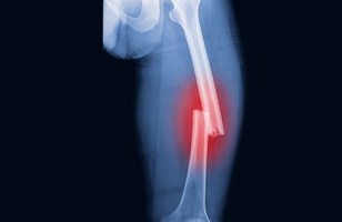
The decreased absorption of phosphate due to chelation with aluminum may cause osteomalacia, or a marked weakening of the bones. As phosphate is necessary for bone calcification, this process occurs at a slower than normal rate. Clinically, this may present as decreased calcification, decreased bone density, or fractures.

Neurons

Some studies have linked high levels of aluminum to Alzheimer’s disease, although this relationship is not causal nor reliably replicated. Alzheimer’s disease is complex, with many different factors affecting the outcome. In animal models, decreased cognitive and motor functions have been noted. Patients on hemodialysis for long periods of time have exhibited signs of neurologic impairment. Some studies of aluminum workers indicated decreased function on nervous function tests.

Lungs

Aluminum workers who inhale larger amounts of aluminum dust may present with signs or symptoms of pulmonary distress. This may include coughing, wheezing, shortness of breath, or pulmonary fibrosis.

Overall, the toxic effects of aluminum appear to be far more prevalent in those with progressive chronic kidney disease. This may be due either to the decreased urinary elimination of aluminum in these patients or, in certain cases, excess aluminum exposure through hemodialysis. Most notably, the bone mineral effects caused by chronic kidney disease may be exacerbated by the bone toxicity of aluminum. Brain disease has been specifically noted in children with kidney diseases.

Management of Poisoned Patients

The most common method for management of aluminum toxicity is avoidance. Decreasing exposure to aluminum containing products such as cosmetics, foods, antacids (aluminum hydroxide), phosphate binders, dialysates, and parenteral injections is recommended. As described above, citrate drastically increases the absorption of aluminum, and thus co-administration of these products should be avoided. Chelating agents such as desferrioxamine (DFO) may be used to chelate aluminum in order to be removed via urine or hemodialysis. This can be especially useful if aluminum deposits are present in the bone (shown by bone biopsy). In rat models, administration of DFO drastically decreases the half-life of aluminum from 150 days to 55 days.

Historical Exposures

One historical exposure of note took place in England in 1989. During this incident, an unknown level of aluminum sulfate was added to drinking water, leading to neurologic symptoms in 5 documented children. These symptoms included memory loss, fatigue, depression, behavioral changes, and learning impairment. However, the validity of this exposure is called into question due to the high levels of copper and lead, known neurotoxins. Other historical exposures have occurred in aluminum-dust-exposed workers as described in the Mechanisms of Action section above.


Conclusion

Overall, aluminum toxicity is very poorly described and the mechanisms still require further elucidation. Aluminum appears to have clear toxic effects in bones, although the pulmonary and neurologic effects require more study.


References

  1. Agency for Toxic Substances and Disease Registry (ATSDR). 2008. Toxicological profile for Aluminum. Atlanta, GA: U.S. Department of Health and Human Services, Public Health Service.
  2. National Center for Biotechnology Information. PubChem Compound Summary for CID 5359268, Aluminum. https://pubchem.ncbi.nlm.nih.gov/compound/Aluminum. Accessed June 14, 2021.
  3. Crisponi, Guido & Fanni, Daniela & Gerosa, Clara & Nemolato, Sonia & Nurchi, Valeria & Crespo-Alonso, Miriam & Lachowicz, Joanna & Faa, Gavino. (2013). The meaning of aluminium exposure on human health and aluminium-related diseases. Biomolecular concepts. 4. 77-87. 10.1515/bmc-2012-0045.
  4. Attitude Pictures Ltd. Vaccines: Aluminium Demonstration [Video]. YouTube. https://www.youtube.com/watch?v=_24rPCn-5PQ. Published August 6, 2018. Accessed June 14, 2021.
  5. MedLecturesMadeEasy. Metabolic Bone Disorders [Video]. YouTube. https://www.youtube.com/watch?v=dmkuUXOZ4EQ&t=328s. Published August 28, 2016. Accessed June 14, 2021.

Brodifacoum Toxicology

Brodifacoum and Superwarfarin Summary

Image 1 Source

Brodifacoum is a second-generation (single-dose) warfarin/4-hydroxycoumarin derivative introduced in 1975 that is commonly used as a potent rodenticide around the world. Brodifacoum’s structure (shown above), is very similar to that of Warfarin (shown in Image 2 below), with which it shares the coumarin moiety. Important differences include the addition of a large, lipophilic, bromine-containing side chain.

Image 2 Source

Brodifacoum and other warfarin derivatives all exert their mechanism of toxicity in the liver mainly through inhibition of vitamin k epoxide reductase, an enzyme that is crucial in the recycling of vitamin k to produce mature clotting factors.  Two critical clotting factors in the common pathway that are reduced are Factors X and II. Figure 1 below shows all factors that are vitamin k-dependent (labeled red) as well as mechanism of action of warfarin derivatives (part B).

Figure 1 Source

According to the available information, brodifacoum does not appear to possess carcinogenic or teratogenic effects (source). This difference in teratogenicity from warfarin may be due to the drastically higher molecular weight, making crossing the barrier of the placenta far more difficult. Brodifacoum’s high lipophilicity lends itself to a high potential for accumulation and extremely long elimination half-life of 56 days (source).


Toxicokinetics and Metabolism

Image 3 Source

While brodifacoum and other “superwarfarins” are similar to warfarin in their high oral toxicity, brodifacoum is also highly toxic when inhaled or absorbed dermally. Brodifacoum is also a minor direct eye and skin irritant. Routes of toxicity for anticoagulant rodenticides are described by the following table:

Table 1. ACUTE TOXICITY CLASSIFICATION – RODENTICIDES
Oral Inhalation Dermal Primary Eye Irritation Primary Skin Irritation
Warfarin3,6 Moderate – High toxicity Not significant Not significant No data No data
Chlorphacinone4 High toxicity High toxicity High toxicity Non-irritating Non-irritating
Diphacinone4 High toxicity High toxicity High toxicity Moderate irritation Slight irritation
Bromadiolone4 High toxicity High toxicity High toxicity Low irritation Minimally irritating
Difethialone3 High toxicity High toxicity High toxicity Mild irritant Non-irritating
Brodifacoum4 High toxicity High toxicity High toxicity Minor irritation Mild irritant
Bromethalin4 High toxicity High toxicity Moderate toxicity Slight irritation Non-irritating
Cholecalciferol3 High toxicity Very low toxicity Low toxicity No data No data
Zinc phosphide5 High toxicity High toxicity Low toxicity Slight irritation Non-irritating
Strychnine7 High toxicity High toxicity Low toxicity Highly irritating Non-irritating
Classification categories were modeled after the U.S. Environmental Protection Agency, Office of Pesticide Programs, Label Review Manual, Chapter 7: Precautionary Labeling. http://www.epa.gov/oppfead1/labeling/lrm/chap-07.pdf

Table 1 Source

As described in detail above, once absorbed, brodifacoum moves to the site of action in the liver. Brodifacoum binds to and inhibits vitamin k epoxide reductase, decreasing the recycling of vitamin k, decreasing the production of mature clotting factors (notably factors X and II), and producing an anticoagulant effect that is around 100 times greater than warfarin (source). Of note, this effect does not happen immediately. Like other coumarin-derived products and drugs, it takes about 3-5 days to for the full effects of the vitamin k antagonist to be seen clinically. Other important pharmacokinetic parameters are as follows (source):

  • MW = 523.4 g/mol
  • LogP = 8.5
  • pKa = 4.5
  • IC 50 = 0.15 μM (Warfarin IC 50 = 2.2 μM)
  • LD 50 (oral) = 0.3 mg/kg (Warfarin in rat models LD 50 = 1.6 mg/kg oral)

While studies of brodifacoum in humans are lacking compared to warfarin, brodifacoum’s mechanism of action and metabolism may subject its effects to similar genetic variations. Of note, warfarin and brodifacoum both inhibit the vitamin k epoxide reductase enzyme (VKOR), which is encoded by the VKORC1 gene in humans. Humans with a mutation causing decrease in function of VKORC1 produce less clotting factors at baseline, thus making them more susceptible to lower doses of warfarin. As brodifacoum shares this same mechanism of action, it is reasonable to believe that those with VKORC1 decreased function mutations may also have an increased anticoagulant response to brodifacoum.

Figure 2 Source

Brodifacoum has shown to be metabolized by multiple pathways, including glucuronidation of the 4-hydroxy group and hydroxylation of the coumarin moiety via hepatic CYP450. As this coumarin system is maintained in both warfarin and brodifacoum, decreases in CYP2C9 function with the *3 allele that cause increased warfarin effect may also cause increased brodifacoum effect (although this has yet to be proven by studies).


Symptoms of Toxicity and Clinical Implications

Image 4 Source

As shown by the simple graphic above, the main clinical symptoms of toxicity are signs of bleeding. These include, but are not limited to:

  • Unusual bruising
  • Bruises that do not heal or continue to get larger
  • Nose bleeds
  • Cuts/abrasions that do not stop bleeding
  • Blood in the urine or stool
  • Blood in the vomit
  • Coughing/vomiting anything resembling coffee grounds
  • Dark, tarry stools
  • Bleeding gums

The effects of brodifacoum and other coumadin-like toxins can be monitored through their effects on the Prothrombin Time (PT), which is normally reported as an International Normalized Ratio (INR). While PT times can vary depending on lab and reagents, a normal INR is around 1-1.1.

Image 5 Source

Most reported pediatric cases of superwarfarin poisoning are through unintentional oral exposure to the pesticide. However, most superwarfarin poisonings in adults are due to attempted suicide and occupational exposure in addition to accidental ingestion (source). Recenlty, in 2018, synthetic cannabis tainted with brodifacoum lead to a large number of cases of coagulopathy and 7 deaths. Due to its identical mechanism of action as warfarin, brodifacoum and other “superwarfarins” can be managed with the same treatments of fresh frozen plasma and vitamin K1 supplementation. Both therapies serve to replenish the absent or drastically decreased clotting factors, with fresh frozen plasma being far more rapid.

With the newest generation of single-dose “superwarfarins” replacing the older and greater-resistance warfarin, it is crucial to be aware of the products containing this rodenticide as well as the signs/symptoms of toxicity these products may produce.


References

  1. National Center for Biotechnology Information. PubChem Compound Summary for CID 54680676, Brodifacoum  https://pubchem.ncbi.nlm.nih.gov/compound/Brodifacoum. Accessed May 30, 2021.
  2. National Center for Biotechnology Information. PubChem Compound Summary for CID 54678486, Warfarin. https://pubchem.ncbi.nlm.nih.gov/compound/Warfarin. Accessed May 30, 2021.
  3. Van Gorp RH, Schurgers LJ. New Insights into the Pros and Cons of the Clinical Use of Vitamin K Antagonists (VKAs) Versus Direct Oral Anticoagulants (DOACs). Nutrients. 2015; 7(11):9538-9557. https://doi.org/10.3390/nu7115479
  4. Rodenticide Products for Consumers; U.S. Environmental Protection Agency, Office of Prevention, Pesticides, and Toxic Substances, Office of Pesticide Programs. http://www.epa.gov/pesticides/reregistration/rodenticides/consumer-prod.html (accessed June 2011), updated June 2011.
  5. Chong Y-K, Mai TW-L. Superwarfarin (Long-Acting Anti-coagulant Rodenticides) Poisoning: from Pathophysiology to Laboratory-Guided Clinical Management. Clinical Biochemist Reviews. 2019;40(4):175-185. doi:10.33176/aacb-19-00029
  6. M. Turner, M. Bula, M. Pirmohamed. Chapter 36 – Personalized Medicine in Cardiovascular Disease, Editor(s): William B. Coleman, Gregory J. Tsongalis. Diagnostic Molecular Pathology, Academic Press, 2017, Pages 457-471, ISBN 9780128008867, https://doi.org/10.1016/B978-0-12-800886-7.00036-4