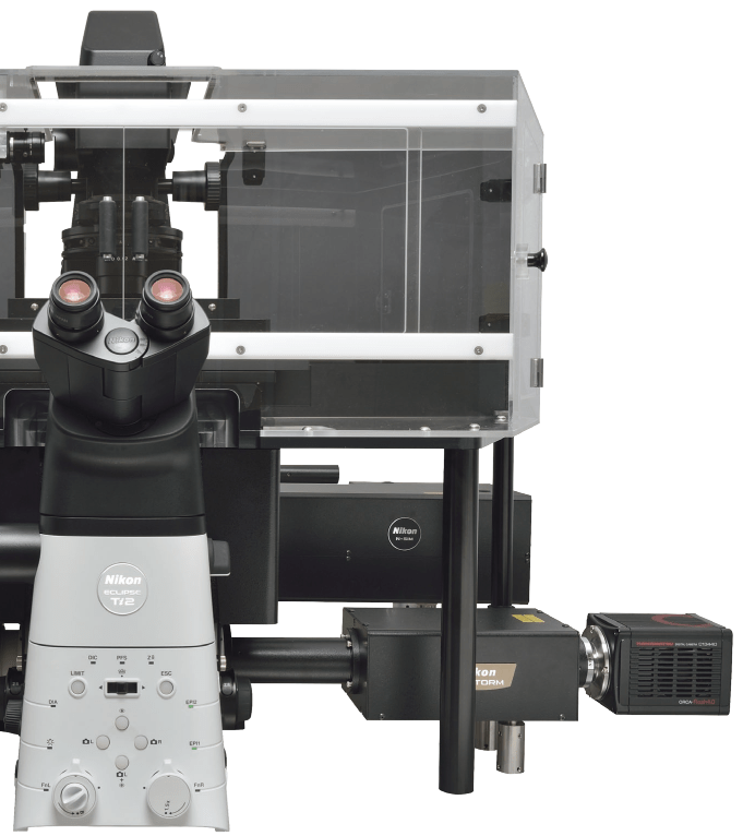
 The Campus Microscopy & Imaging Facility (CMIF) at the Ohio State University has recently acquired a new high resolution microscope that uses the latest in high resolution fluorescence techniques. The new system is from Nikon, called the N-SIM S. It includes integrated components to perform super-resolution structured illumination microscopy (SIM) and stochastic optical reconstruction microscopy (STORM). This post highlights four examples of what the system can do and briefly describes the basic principles behind the underlying microscopy techniques.
Examples of the Nikon N-SIM S in Action
Our Nikon N-SIM S microscope system has several imaging modes: 3D-SIM, TIRF-SIM, and STORM. Below are some examples of what the Nikon N-SIM S system can do in these different modes.
Below is an image of microtubules within a melanoma cell taken with the N-SIM S in 3D-SIM mode. The 3D-SIM image is juxtaposed with an image of the same cell with the same microscope in conventional widefield mode. The 3D-SIM image has less background and greater contrast than the conventional widefield image. The 3D-SIM is capable of lateral (xy) resolutions as low as 115 nm.

Microtubules shown in an image taken by the Nikon N-SIM S in 3D-SIM mode (left) compared to an image in conventional widefield mode (right). (Source: Nikon)
The Nikon N-SIM S also has high resolution in the axial (z) direction. Below are 3D projections of dendritic spines. One taken with the N-SIM S in 3D-SIM mode and the other taken with the Nikon A1R confocal microscope. The zoomed in image at the bottom of each image shows that the N-SIM S is able to achieve greater resolution in the z-axis compared to the A1R confocal microscope. The N-SIM S in 3D-SIM mode is capable of achieving resolutions in the axial direction down to 269 nm.

Dendritic spines shown in 3D projections taken by the Nikon N-SIM S in 3D-SIM mode (left) compared to an image taken by the Nikon A1R confocal microscope (right). (Source: Nikon)
In addition to the 3D-SIM mode, the Nikon N-SIM S has a mode that uses total internal reflection fluorescence (TIRF) combined with SIM, called TIRF-SIM. Below are images of the plasma membrane of a melanoma cell. An image, taken in TIRF-SIM mode, is compared to an image taken with a conventional TIRF microscope. The TIRF-SIM achieves dramatically higher resolutions than a conventional TIRF. The TIRF-SIM mode is capable of achieving lateral resolutions down to 86 nm.

A TIRF-SIM image (left) compared to a conventional TIRF image (right) of the plasma membrane of a melanoma cell. (Source: Nikon)
In addition, the Nikon N-SIM S can capture fast processes within live cells. Below is an image from a time series, taken at 5 frames per second, with the N-SIM S in 3D-SIM mode. The time series shows the movement of endosomes within a live cell. The N-SIM S is capable of image acquisition speeds of up to 15 frames per second.

Endosomes within a live cell imaged with the Nikon N-SIM S in 3D-SIM mode (left and middle), and a widefield image shown for comparison (right). Click the image to see the time series. (Source: Nikon)
Even greater resolutions are achievable with the Nikon N-SIM S microscope when in N-STORM mode. This mode uses the super resolution technique called stochastic optical reconstruction microscopy (STORM). Below is an image of subcellular components of a kidney cell with the alpha-tubulin (magenta), the caveolin (red), and the f-actin (green) labeled. In N-STORM mode, the N-SIM S is capable of achieving down to 20 nm lateral resolution and down to 50 nm axial resolution.

An N-STORM image showing fluorescently labeled alpha-tubulin (magneta), caveolin (red), and f-actin (green) within a kidney cell. (Source: Nikon)
For more information about the Nikon N-SIM S microscope system and what it can do, visit the Nikon website.
Background
The Nikon N-SIM S microscope uses multiple high resolution microscopy techniques. These include structured illumination microscopy (SIM), total internal reflection microscopy (TIRF), and stochastic optical reconstruction microscopy (STORM). The basic principles behind each technique is described below.
Structured Illumination Microscopy (SIM)
Structure illumination microscopy (SIM) is a technique involving some interesting but complex mathematics, which are beyond the scope of this article. However, I will briefly describe the basic process by which SIM creates high resolution images. When laser light with a high frequency pattern illuminates a sample, it interferes with the high frequency information in the sample. This high frequency information in the sample is usually hidden. However, the light pattern interferes with the high-frequency information in the sample and reveals the hidden information. The unveiling of this information takes the form of an interference pattern, called a Moire pattern. Several Moire patterns are collected with the light patterns at several different orientations and then computationally combined to create a high resolution image.

Moire Effect. When a sample (a) is illuminated with light with high frequency patterns (b), the high frequency patterns in the sample combine with the patterns in the light and create interference patterns (c), revealing high frequency information in the sample. (Source: Gustafsson, 2005.)
For more information on super-resolution structured illumination microscopy, see this article or the review by Heintzmann and Huser (2017).
Total Internal Reflection Fluorescence (TIRF) Microscopy
The Nikon N-SIM S microscopy system combines SIM with total internal reflection fluorescence (TIRF) microscopy to achieve higher resolution images above SIM alone. TIRF works by using a fascinating electromagnetic effect that occurs when light reflects off of a surface under specific conditions. When laser light is directed at a glass slide, some of the light passes through the slide and some is reflected back. As the angle of the laser light is increased, more and more of the light is reflected back. When the laser light reaches a specific angle of reflection off the inside of the glass slide, the laser light is completely reflected. Fascinatingly, an evanescent wave of light is created on the surface of the glass slide. This wave excites fluorophores within the sample just ~100 nm from the glass slide surface. This confinement of the fluorophore excitation leads to high resolution images.

TIRF. The creation of an evanescent wave at the surface of glass slide by laser light at a glancing angle activates fluorophores only ~100 nm within a cell. (Source: Molecular Expressions)
For more information on total internal reflection microscopy, see this article at MicroscopyU.com.
Stochastic Optical Reconstruction Microscopy (STORM)
Stochastic optical reconstruction microscopy (STORM) takes advantage of the optical properties of fluorescent dyes to obtain high resolution images. First, the fluorophores are induced into a reversible dark state (i.e., non-fluorescent). Then low-power laser light is directed onto the sample, exciting only a tiny subset of fluorophores in a statistically random manner. The fluorophores are imaged, and then the center point for each fluorophore is determined computationally. By imaging with a low-power laser over a long enough duration, centers for all of the fluorophores can be determined individually, and then an image is reconstructed from the center points. Through this process, the STORM technique is able to achieve high resolution images.

STORM. Unlike conventional fluorescence microscopy, STORM excites only a tiny subset of fluorophores at a time (red = emitting fluorescence; gray = dark). The signal from which can be used to determine computationally the center points of the individual fluorophores (crosses). After collecting signals from every fluorophore, the center points are used to reconstruct an image with higher resolution than conventional fluorescence microscopy. (Source: The Vaughn Group at the University of Washington)
For more information on stochastic optical reconstruction microscopy, see this article at MicroscopyU.com.
CMIF Capabilities
We have acquired the Nikon N-SIM S super resolution microscope including the N-STORM system. We are currently remodeling the CMIF to accommodate the new microscope system. Please check the CMIF website for latest details on the availability of the system.
In the meantime, the CMIF currently has available for use the Nikon A1R live cell microscope system. This confocal microscope is great for imaging live cells. For more information about the Nikon A1R live cell microscope system at CMIF can be found on the CMIF website.
References
“Nikon Small World image competition.” www.nikonsmallworld.com. Nikon, Web. 27 Apr. 2020.
“N-SIM S microscope system.” www.microscope.healthcare.nikon.com. Nikon, Web. 27 Apr. 2020.
“N-STORM microscope system.” www.microscope.healthcare.nikon.com. Nikon, Web. 27 Apr. 2020.
“Molecular Expressions. Exploring the world of optics and microscopy.” micro.magnet.fsu.edu. Florida State University, Web. 29 Apr. 2020.
Demmerle, J. “A semi-intelligible explanation of structured illumination microscopy (SIM).” BiteSizeBio.com. Web. 1 May 2020.
Gustafsson, M.G.L. 2005. “Nonlinear structured-illumination microscopy: wide-field fluorescence imaging with theoretically unlimited resolution.” PNAS, vol. 102, pp. 13081-13086.
Ross S.T., S. Schwartz, T.J. Fellers, and M.W. Davidson. “Total Internal Reflection Fluorescence (TIRF) Microscopy.” MicroscopyU.com. Nikon, Web. 1 May 2020.
The Vaughn Group. STORM. https://sites.google.com/a/uw.edu/the-vaughan-group/storm. University of Washington, Web. 29 Apr. 2020.
Heintzmann, R. and T. Huser. 2017. “Super-resolution structured illumination microscopy.” Chemical Reviews, vol. 117, pp. 13890-13908.
Allen, J.R., J.S. Silfies, S.A. Schwartz, and M.W. Davidson. “Single-molecule super-resolution image.” MicroscopyU.com. Nikon, Web. 1 May 2020.