Medical Knowledge and Skills CEO 2.
The graduate is able to:
- demonstrate a broad working knowledge of the fundamental science, principles, and processes basic to the practice of medicine and apply this knowledge in a judicious and consistent manner to prevent common health problems and achieve effective and safe patient care.
- understand the clinical relevance of scientific inquiry and demonstrate the ability to evaluate emerging knowledge and research as it applies to diagnosis, treatment and the prevention of disease.
- utilize state of the art information technology and tools to retrieve, manage and use biomedical information in the care of individuals and populations.
- understand the indications, contraindications, and potential complications of common clinical procedures and perform the basic clinical procedures expected of a new PGY-1
Ultrasound as a technology is developing more and more uses in medicine. Being an inexpensive, nonionizing imaging technology makes it attractive for many applications. At Ohio State, I am fortunate to be part of one of the best medical student ultrasound curriculums in the country. I remember being enthralled by Dr. Bahner’s first lecture on ultrasound during my first year of medical school. I promptly signed up as a trained simulated ultrasound patient (TSUP) and for what was then called Trinity Ultrasound (now called Beginner Ultrasound). I modelled my way through my first Ultrafest (i.e., OSU’s regional conference on ultrasound) during Snowmageddon. I learned how the FAST scan had largely supplanted the diagnostic peritoneal lavage. (In my fourth year, I did see two SICU attendings speculating how they might need to do a diagnostic peritoneal lavage. They reminisced about how that procedure is barely taught these days.) I had my eye scanned and watched my consensual pupillary response. This ultrasound technique is more than a parlor trick. In a patient who cannot open one eye due to swelling, ultrasound can check if the consensual pupillary response is intact.
In my second year, I kept modelling and moved on to Intermediate Ultrasound. I competed in Ultrafest that year. My teammate was a randomly assigned first year medical student. Other teams had four fourth years. We put our hearts into it, but we came up against a game where ultrasound images were covered by rectangles that slowly fell away. The first team to buzz in was able to answer. Two against several teams of four, we didn’t make it any farther. But that really wasn’t the point – what really hit home was just how amazing this technology could be in practice. At the new James Cancer Hospital and Solove Research Institute, I learned how ultrasound was being used to place brachytherapy. At an interventional radiology interest group meeting, I learned how to perform fine needle aspiration, Figure 1. Later, in my fourth year, I would teach that technique to second year medical students.
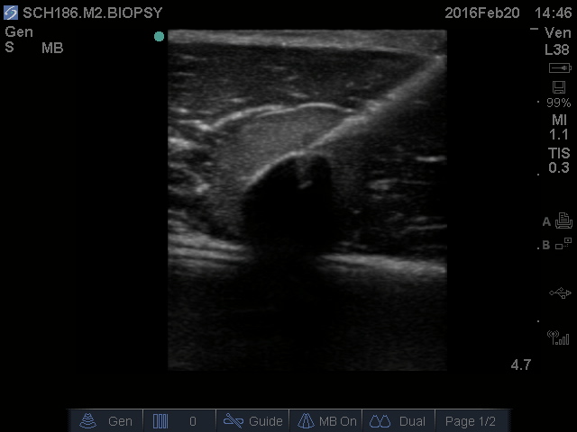
Figure 1: Fine needle aspiration.
In my third year, I moved on to Advanced Ultrasound, and started to proctor sessions, teaching what I had learned. It is often said that ultrasound imaging is operator dependent, but what isn’t often said that training basic ultrasound competency is relatively easy. There are multiple papers that describe what happen when residents with minimal ultrasound training are sent off to perform diagnostic ultrasounds. These papers report high sensitivity and specificity for all manners of conditions, including sepsis. In another example, Dr. Blankenship developed the Army’s First Tactical Ultrasound Course in 2007. In order to convince the military brass that ultrasound was teachable, he taught it to cooks (food preparation specialists) and showed their results were accurate. In teaching ultrasound to medical students, I do my part to make ultrasound understandable and reproducible.
On one of my 24 hour calls, I put what I had learned into action by performing a FAST scan on a trauma patient who had fallen off a ladder. His FAST was negative. I settled a point of contention between a fellow and a resident using ultrasound. The fellow was convinced a post-op mastectomy patient had a seroma. The resident was convinced she did not. I suggested to the resident that if we could find a ultrasound machine, we could take a look. Acquiring an ultrasound machine proved a problem. We ended up needing to consult the PICC team so we could borrow their ultrasound machine. The imaging very clearly showed that the patient had no seroma. Her tissue was simply swollen and inflamed. We had no way of saving the image off the PICC team machine, so the resident took a de-identified cell phone photo and showed it to the fellow.
Honors Ultrasound ushered in the start of my fourth year of medical school. I designed a pin for the Ultrasound Interest group, Figure 2. As mentioned previously, for my Honors Ultrasound project, I taught thyroid ultrasound to the second year medical students, teaching fine needle aspiration, general thyroid anatomy, volume measurement, locating the superior thyroid artery, and watching the vocal cords with ultrasound. I blunt dissected pockets into chicken breast and hid objects inside to give the student something to sample with fine needle aspiration, Figure 3.

Figure 2: Curvilinear ultrasound transducer pin.

Figure 3: Suspicious object.
At Ultrafest, I staffed the beginner track registration with Yalan. We checked in students for so long that breakfast ran out before we could get there. There wasn’t even coffee left. Then I proctored head and neck ultrasound in the morning, Figure 4. Next was the scavenger hunt, and I was pleased to see that some of my thyroid ultrasound students had retained knowledge from my session the week prior. I didn’t have a model for my station, so I just let students scan on me while checking off the structures they were able to find on their scavenger hunt list. Finally, I walked students through how to perform pericardiocentesis on a phantom. Sammie King brought me my first coffee of the day, earning herself medical school sainthood.

Figure 4: “Baby back rib” sign of tracheal rings.
In the SICU, a resident and I used ultrasound to place a tricky femoral line, Figure 5. We later did a FAST on a patient who had taken in liters of fluid, had minimal urine output, and no edema on physical exam. He didn’t have any intraperitoneal fluid, which lessened our concern about a bleed and raised intracellular shift of fluid on our differential.

Figure 5: Femoral artery showing brisk arterial waveform.
On my endocrine rotation, I saw many thyroid nodules and fine needle aspirations in action. As a resident, I will continue learning appropriate applications of ultrasound. Ultrasound will be a lifelong skill I will use in patient care. I’m lucky I was able to go to OSUCOM, with is strong history of medical student training, and I will keep passing on what I have learned in the tradition of medical education.
[cite papers at the end]