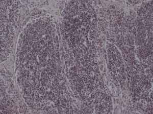
Histology: the study of the microscopic structure of tissues
I took the above images as part of a project completed through my introductory animal science course. My group chose to study the mammary gland, or udder, of a dairy cow. Throughout the semester, we collected tissue samples from various parts of the mammary gland, prepared and preserved these tissue samples in paraffin wax, and sectioned off thin slices of the tissue samples using a microtome. Then we positioned each sample on a microscope slide and stained them. Lastly, after much research, we viewed our slides under a microscope and took various photographs. As a culmination of the project, we presented our photographs and research to the rest of the class at the end of the term.
The goal of this project was to connect the microscopic form of each tissue sample with the gross function of the living organ. By completing this project, I not only learned a great deal about mammary gland form and function but also gained invaluable experience in a laboratory. This project made me much more comfortable in a lab setting using lab equipment. Although it was a lot of hard work, this project also gave me something of which to be proud and I have extremely thankful to have been given such an amazing opportunity.

