Population pharmacokinetics of lenalidomide in patients with B-cell malignancies

Aims: Lenalidomide is an immunomodulatory imide drug used broadly in the treatment of multiple myeloma and lymphoma. It continues to be evaluated in chronic lymphocytic leukaemia (CLL) at lower doses due to dose-related toxicities including tumour flare and tumour lysis syndrome. This study aimed to develop a population pharmacokinetic model for lenalidomide in multiple cancers, including CLL, to identify any disease-related differences in disposition.

Methods: Lenalidomide concentrations from 4 clinical trials were collated (1999 samples, 125 subjects), covering 4 cancers (multiple myeloma, CLL, acute myeloid leukaemia and acute lymphoblastic leukaemia) and a large dose range (2.5-75 mg). A population pharmacokinetic model was developed with NONMEM and patient demographics were tested as covariates.

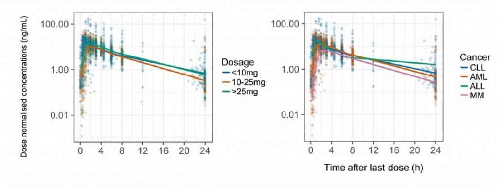
Results: The data were best fitted by a 1-compartment kinetic model with absorption described by 7 transit compartments. Clearance and volume of distribution were allometrically scaled for fat-free mass. The population parameter estimates for apparent clearance, apparent volume of distribution and transit rate constant were 12 L/h (10.8-13.6), 68.8 L (61.8-76.3), and 13.5 h-1 (11.9-36.8) respectively. Patients with impaired renal function (creatinine clearance <30 mL/min) exhibited a 22% reduction in lenalidomide clearance compared to patients with creatinine clearance of 90 mL/min. Cancer type had no discernible effect on lenalidomide disposition.

Conclusions: This is the first report of a lenalidomide population pharmacokinetic model to evaluate lenalidomide pharmacokinetics in patients with CLL and compare its pharmacokinetics with other B-cell malignancies. As no differences in pharmacokinetics were found between the observed cancer-types, the unique toxicities observed in CLL may be due to disease-specific pharmacodynamics.

Figure: Plasma lenalidomide concentration–time profiles normalised for dose. The coloured dots represent each data point, with the colours corresponding to the dose ranges A, or cancer type B, according to the legend. ALL, acute lymphoblastic leukaemia; AML, acute myeloid leukaemia; CLL, chronic lymphocytic leukaemia; MM, multiple myeloma.

Hughes JH, Phelps MA, Upton RN, Reuter SE, Gao Y, Byrd JC, Grever MR, Hofmeister CC, Marcucci G, Blum W, Blum KA, Foster DJR. Population pharmacokinetics of lenalidomide in patients with B-cell malignancies. Br J Clin Pharmacol. 2019 May;85(5):924-934. doi: 10.1111/bcp.13873. Epub 2019 Feb 27. PMID: 30672004; PMCID: PMC6475687.

Pharmacokinetic-Pharmacodynamic Model of Neutropenia in Patients With Myeloma Receiving High-Dose Melphalan for Autologous Stem Cell Transplant

High-dose melphalan (HDM) is part of the conditioning regimen in patients with multiple myeloma (MM) receiving autologous stem cell transplantation (ASCT). However, individual sensitivity to melphalan varies, and many patients experience severe toxicities. Prolonged severe neutropenia is one of the most severe toxicities and contributes to potentially life-threatening infections and failure of ASCT. Granulocyte-colony stimulating factor (G-CSF) is given to stimulate neutrophil proliferation after melphalan administration. The aim of this study was to develop a population pharmacokinetic/pharmacodynamic (PK/PD) model capable of predicting neutrophil kinetics in individual patients with MM undergoing ASCT with high-dose melphalan and G-CSF administration. The extended PK/PD model incorporated several covariates, including G-CSF regimen, stem cell dose, hematocrit, sex, creatinine clearance, p53 fold change, and race. The resulting model explained portions of interindividual variability in melphalan exposure, therapeutic effect, and feedback regulation of G-CSF on neutrophils, thus enabling simulation of various doses and prediction of neutropenia duration.

Visual predictive check (VPC) plot of the final model simulated data vs. observed data in (a) all patients, (b) with granulocyte‐colony stimulating factor (G‐CSF) regimen starting on day +1, and (c) with G‐CSF regimen starting on day +7 after transplantation. Blue dots, the observed data; black dashed line, 2.5th and 97.5th percentiles of the observed data; black solid line, the median of the observed data; red solid line, the median of the simulated data; gray area, 95% prediction interval of the simulated data; black dashed straight line, absolute neutrophil count (ANC) = 0.5 K cells/μL.
Figure: Visual predictive check (VPC) plot of the final model simulated data vs. observed data in (a) all patients, (b) with granulocyte‐colony stimulating factor (G‐CSF) regimen starting on day +1, and (c) with G‐CSF regimen starting on day +7 after transplantation. Blue dots, the observed data; black dashed line, 2.5th and 97.5th percentiles of the observed data; black solid line, the median of the observed data; red solid line, the median of the simulated data; gray area, 95% prediction interval of the simulated data; black dashed straight line, absolute neutrophil count (ANC) = 0.5 K cells/μL.

Cho YK, Irby DJ, Li J, Sborov DW, Mould DR, Badawi M, Dauki A, Lamprecht M, Rosko AE, Fernandez S, Hade EM, Hofmeister CC, Poi M, Phelps MA. Pharmacokinetic-Pharmacodynamic Model of Neutropenia in Patients With Myeloma Receiving High-Dose Melphalan for Autologous Stem Cell Transplant. CPT Pharmacometrics Syst Pharmacol. 2018 Nov;7(11):748-758. doi: 10.1002/psp4.12345. Epub 2018 Oct 20. PMID: 30343510; PMCID: PMC6263666.

Cachectic Cancer Patients: Immune to Checkpoint Inhibitor Therapy?

Immune checkpoint inhibition is dramatically improving patient outcomes in diverse cancers, many of which responded poorly to traditional cytotoxic agents. Drivers of heterogeneous response to immune checkpoint therapy are poorly characterized. Cachectic cancer patients exhibit elevated pembrolizumab clearance and poor response, highlighting the immense therapeutic challenge posed by cancer cachexia. See related article by Turner et al., p. 5841.

Figure 1: Cachexia/malnutrition blocks the beneficial effects of pembrolizumab independent of pembrolizumab exposure.

Coss CC, Clinton SK, Phelps MA. Cachectic Cancer Patients: Immune to Checkpoint Inhibitor Therapy? Clin Cancer Res. 2018 Dec 1;24(23):5787-5789. doi: 10.1158/1078-0432.CCR-18-1847. Epub 2018 Jul 17. PMID: 30018117; PMCID: PMC6279566.

Comprehensive toxicity and immunogenicity studies reveal minimal effects in mice following sustained dosing of extracellular vesicles derived from HEK293T cells

Extracellular vesicles (EVs) are under evaluation as therapeutics or as vehicles for drug delivery. Preclinical studies of EVs often use mice or other animal models to assess efficacy and disposition. However, as most EVs under evaluation are derived from human cells, they may elicit immune responses which may contribute to toxicities or enhanced EV clearance. Furthermore, EVs from different cell sources or EVs comprising various cargo may differ with respect to immunogenicity or toxicity. To assess EV-induced immune response and toxicity, we dosed C57BL/6 mice with EVs intravenously and intraperitoneally for 3 weeks. EVs were harvested from wild type or engineered HEK293T cells which were modified to produce EVs loaded with miR-199a-3p and chimeric proteins. Blood was collected to assess hematology, blood chemistry, and immune markers. Spleen cells were immunophenotyped, and tissues were harvested for gross necropsy and histopathological examination. No signs of toxicity were observed, and minimal evidence of changes in immune markers were noted in mice dosed with engineered, but not with wild type EVs. This study provides a framework for assessment of immunogenicity and toxicity that will be required as EVs from varying cell sources are tested within numerous animal models and eventually in humans.

Measurements of different cell populations in spleen cells by flow cytometry using appropriate surface markers conjugated with different fluorochromes. Fluorescence signal of T-cell surface marker CD3e conjugated with PE-Vio770 (P1 = T cells) and B-cell surface marker CD19 conjugated with APC-Vio770 (P2 = B cells) on the cells from mice receiving (a) vehicle control or (b) lipopolysaccharide (LPS). The percentage of each cell population in mice (c) 24 h after LPS treatment (three doses, n = 4) or (d) 3 weeks after treatment with extracellular vesicles (10 doses, n = 10). Bars and error bars denote the mean and standard deviation, respectively, of experimental groups. PBS, phosphate-buffered saline; WT, wild-type HEK293T cells. *p < 0.05.
Figure: Measurements of different cell populations in spleen cells by flow cytometry using appropriate surface markers conjugated with different fluorochromes. Fluorescence signal of T-cell surface marker CD3e conjugated with PE-Vio770 (P1 = T cells) and B-cell surface marker CD19 conjugated with APC-Vio770 (P2 = B cells) on the cells from mice receiving (a) vehicle control or (b) lipopolysaccharide (LPS). The percentage of each cell population in mice (c) 24 h after LPS treatment (three doses, n = 4) or (d) 3 weeks after treatment with extracellular vesicles (10 doses, n = 10). Bars and error bars denote the mean and standard deviation, respectively, of experimental groups. PBS, phosphate-buffered saline; WT, wild-type HEK293T cells. *p < 0.05.

Zhu X, Badawi M, Pomeroy S, Sutaria DS, Xie Z, Baek A, Jiang J, Elgamal OA, Mo X, Perle K, Chalmers J, Schmittgen TD, Phelps MA. Comprehensive toxicity and immunogenicity studies reveal minimal effects in mice following sustained dosing of extracellular vesicles derived from HEK293T cells. J Extracell Vesicles. 2017 Jun 6;6(1):1324730. doi: 10.1080/20013078.2017.1324730. PMID: 28717420; PMCID: PMC5505007.