As a follow-up to my earlier blog post, I thought I should show some more examples or successes and problems in imaging mites.
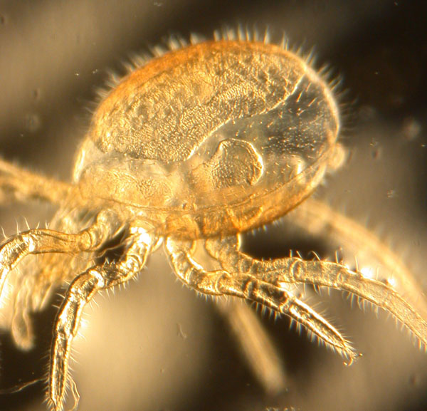
Diplothyrus lecorrei (Holothyrida), nymph, single image using dissecting microscope
The problem in the Diplothyrus image is depth of field. The marginal areas of this mite are out of focus.
The LT-SEM image of Excelsotarsonemus shows what great depth of field can do. Note the blobs of fungus stuck on this mite. They are vectors of specific fungi.

Excelsotarsonemus sp. (Tarsonemidae), colorized LT-SEM
Stacked imaging with Differential Interference Contrast illumination works very well with well sclerotized mites. It generates nice images of cuticular patterns.
-

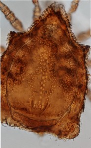
- Heterozerconidae, n.gen., n. sp. (Sejina), stacked image, 200x
-
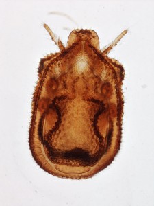
- Macrodinychus sp. (Uropodina), stacked image at low magnification (40x)
-

- Nanhermannia sp. (Oribatida) stacked image, 40x
-

- Micromegistus bakeri (Trigynaspida), female with egg, stacked image, 200x
-
- Eutrachytes sp. (Uropodina), stacked image of dorsum
The technique can work well on poorly sclerotized mites, but in this case one has to worry about avoiding imaging structures from the other side.
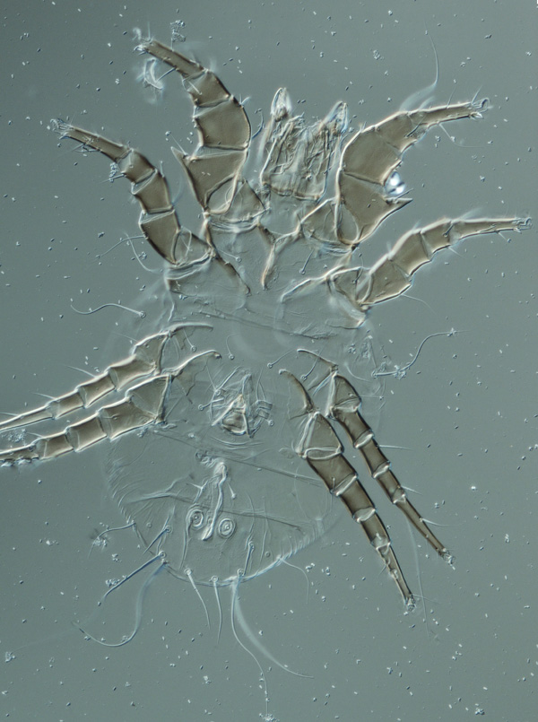
Acarus siro, male, stacked image
The Trichocylliba images are a good example of what happens with stacked images to 3-dimensional structure. These mites are as high as they are long, but the images do not show that. Adding lateral images would solve it, but that proved very tricky with these mites.

Trichocylliba n.sp. (Uropodina), male, stacked image, 200x
Just for fun, a partial slice though the mouthparts of this Oudemansicheyla shows some of the power of Confocal microscopy (image courtesy of Sam Bolton and Gary Bauchan).

Oudemansicheyla (Cheyletidae), confocal microscopy
About the Author: Dr. Hans Klompen is professor in the Department of Evolution, Ecology and Organismal Biology and Director of the Ohio State University Acarology Collection.
Terrific ! Your images are terrific ! Thanks for testing different technics. DIC seems to be a very interesting technic even for dead organisms. Can you tell us the way you clarify the mites and the mounting media ?
What is the type of dissecting microscope you used ?
Many thanks for your splendid images !
MicrOlivier