
What Is It? | Facts in Depth | For the Professional Diagnostician
Brassica Diseases | Black Rot
Black Rot
Symptoms
Seedlings
Seedlings are very susceptible to black rot infection. When infected, seedlings become systemically infected, exhibiting chlorosis on the young leaves. As the disease progresses, seedlings prematurely drop leaves and eventually die.
Mature Plants
On mature plants, leaf edges are chlorotic or necrotic. Lesions form in a distinct “v-shape,” with the base of the “V” directed along the vein. Closer inspection of these infected areas with a hand lens reveals vein discoloration. These veins usually appear brown to black in color, hence the name black rot. Lesions eventually expand downward toward the base of the leaf and may move down the vascular tissue of petioles and then spread up and down the stems. When infected stems and petioles are cut crosswise or lengthwise, vascular tissue is black to brown in color with yellowish bacterial slime. This leads to wilting and eventual death of the plant.


(Left and Middle Pictures: Courteousy of the American Phytopathological Society)
Signs
Under certain environmental conditions, bacteria can ooze from the stems and infected areas. Bacterial streaming also occurs when the lesions are cut marginally, placed in water on a glass slide and under a cover slip, and under a microscope with dark background.


Often Confused With
- Nutrient deficiencies – Nutrient deficiency symptoms in crucifers can resemble those of black rot. For example, a magnesium deficiency in crucifers causes chlorosis on interveinal areas of lower leaves. These chlorotic regions eventually become necrotic.
- Fusarium yellows – Symptoms are very similar to black rot; however, vein discoloration is brown, and bacterial slime is not present.
Isolation Media
- Detection of Xanthomonas campestris pv. campestris on Brassica spp. disinfested/disinfected seed with grinding
- mCS20ABN – After 4–6 d on mCS20ABN, X. campestris pv. campestris colonies are yellow, mucoid and surrounded by a zone of starch hydrolysis.
- FS – After 4–6 d, X. campestris pv. campestris colonies are small, pale green, mucoid and surrounded by a zone of starch hydrolysis. This zone appears as a halo that may be easier to see with a black background.
- Yeast extract-dextrose-calcium carbonate (YDC) medium is a non-selective media used for subculturing suspected xanthomonads and clavibacters. Xanthomonas spp. colonies on YDC medium are yellow, mucoid, round, entire and raised after 3 days at 82 °F.
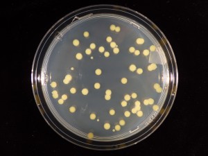
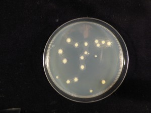

(Courteousy of Dr. Francesca Peduto-Hand at The Ohio State University; Left: CKTM Media; Middle: mCS20ABN Media; Right: YCD Media)
Rapid Diagnostic Tests
- Agdia Immunostrip Test®
- The primers XCF and XCR were designed from hrpF of X. c. pv. campestris, with a predicted PCR product of 535 bp (5′-CGATTCGGCCATGAATGACT-3′ and 5′-CTGTTGATGGTGGTCTGC AA-3′) from the paper Park et. al., 2004.