
Dig deeper ♣ Download fact-sheet
Cytospora Canker of Conifers
by Paige Thrush, Nancy J. Taylor & Francesca Peduto Hand
Identification
Cytospora canker is a stem and branch disease, causing dieback on hundreds of different species of trees and shrubs. The disease is especially destructive to spruces and other conifers in the Midwest and eastern U.S., most often damaging species that are stressed and/or planted outside of their native ranges.Symptoms associated with Cytospora species include discolored foliage, premature defoliation, branch dieback, and cankers. The disease usually affects trees older than 15 years that are or have been stressed by drought, other diseases, or cultural problems. From a distance, the most obvious symptom of the disease includes dead branches, often starting in the lower part of the tree (Figure 1). If conditions are conducive in the following years, the disease will begin to spread upward.
Although canker is in the disease name, this symptom may or may not be readily visible on the host. Cankers first appear as discolored, sunken areas of bark at the base of a stem or branch, usually growing to be elongate or diamond shaped. As the fungus colonizes the vascular tissue of the plant, cankers eventually girdle, or encircle, branches, killing the branch and foliage above the canker site (Figure 2). Small branches can be girdled and killed in one to two years, while it may take several years to decades for the fungus to girdle large branches or trunks. Cankers may go unnoticed for years until the growth of new tissue causes the lesion to appear sunken. Often, a canker cannot be observed until the diseased bark has fallen from the tree or has been cut off to reveal the dead wood underneath (Figure 3).
Another phenomenon that may occur is resin flow, which is caused by a defensive response in the host plant that is especially evident in spruce species (Figure 4). Resin flow is often observed at the perimeter of the diseased tissue, but may drip down onto lower branches where there are actually no cankers. The resin may be a clear or amber color and will eventually crystallize to form a white crust.
Small fungal fruiting bodies may appear on or near the canker. These fruiting bodies hold the spores, or reproductive structures, of the pathogen, which allow it to survive, spread, and cause new infections. To observe these tiny fruiting structures, use a hand lens and look on the diseased canker tissue and in adjacent areas on the bark. During wet periods, yellow to orange spore tendrils may be observed coming out of the bark (Figure 5).
Once a branch or stem has been girdled, symptoms of dieback occur. Needles on infected branches will turn yellow, brown, purplish-brown, or reddish-brown in spring or summer during the first year of infection. Discolored needles often drop off the tree during winter, leaving bare, brittle and dead branches that may remain on the tree for many years. Small branches that have been infected may not produce new needles in spring of the following growing season. The result of Cytospora infections is a disfigured host with a barren and asymmetrical appearance. If disease is suspected, samples can be sent to a diagnostic laboratory for confirmation. Information on The Ohio State University laboratory can be found on the C. Wayne Ellett Plant and Pest Diagnostic Clinic website.
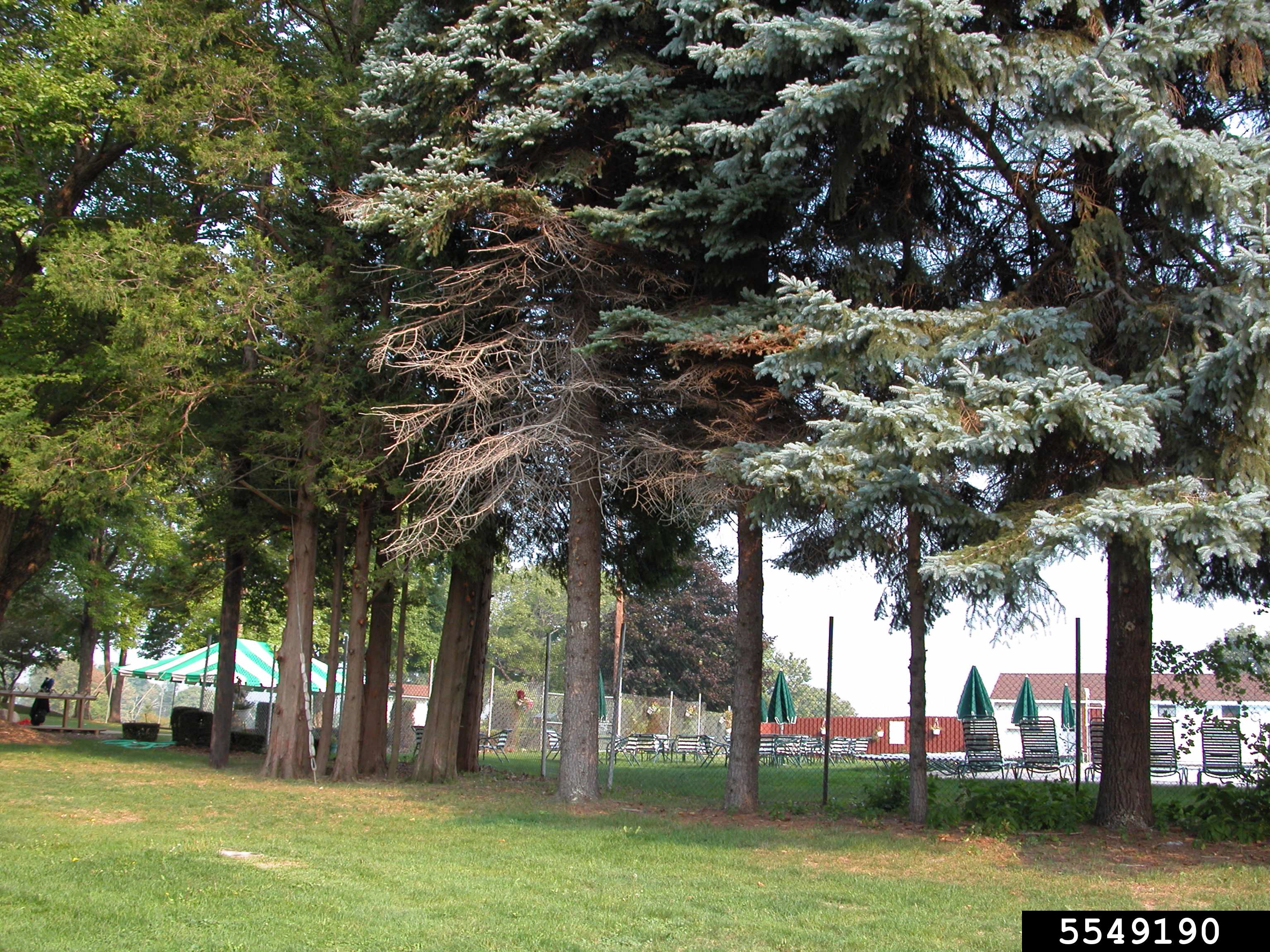

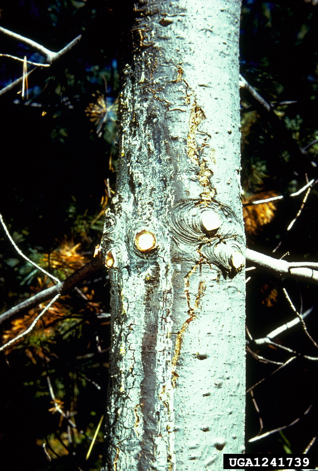
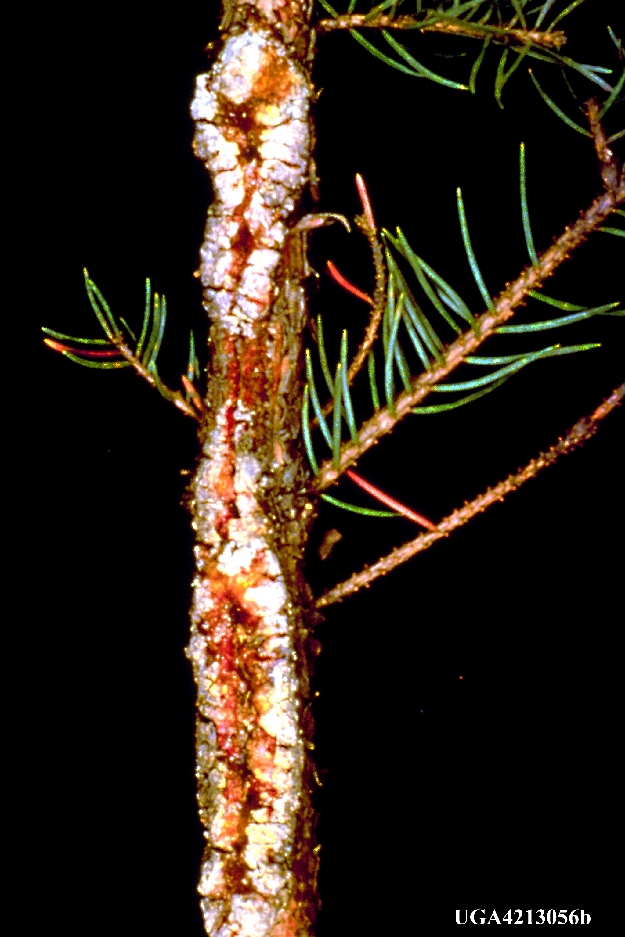

From top left to bottom right: Figure 1. Cytospora canker on the lower branches of a spruce tree (Penn State Department of Plant Pathology & Environmental Microbiology Archives, Penn State University, Bugwood.org); Figure 2. Canker progressing from a branch into the stem (Robert L. James, USDA Forest Service, Bugwood.org); Figure 3. Canker on trunk ad stems of spruce and fir (USDA Forest Service, USDA Forest Service, Bugwood.org) ; Figure 4. Resin flow over branch canker on blue spruce (Minnesota Department of Natural Resources, Minnesota Department of Natural Resources, Bugwood.org); Figure 5. Spore tendrils emerging from the trunk of a tree (William Jacobi, Colorado State University, Bugwood.org).
Management Guidelines
Integrated pest management (IPM) is an approach to plant health care and disease and pest control. IPM incorporates a wide range of strategies to prevent, minimize, and/or control abiotic and biotic diseases and pests. These strategies involve monitoring and scouting, learning behavior and life cycle of pests and pathogens, accurately identifying the source of disease, developing threshold levels, employing preventive measures and integrating cultural, mechanical, biological and chemical controls. All management decisions should be carried out based upon the specific requirements of the plant. Analysis of each strategy involves considering the impact on host plants, pests/pathogens, microbial interactions, the environment and society. The costs and benefits of each strategy should be evaluated before making management decisions.
Prevention:
- Purchase certified pest and pathogen-free stock from reputable sources.
- Nurseries:
- Quarantine incoming plants and inspect for signs and symptoms of disease. Remove and dispose of, or return, all infected plants to the original source.
- Proper site selection:
- Avoid drought prone sites and those that may become waterlogged.
- When planting a young tree in the landscape, consider the future requirements of the mature tree, and select a site that will provide the specimen with adequate space, water, nutrients and sunlight.
- Use proper transplanting techniques to prevent damage to roots.
- Prevent and minimize stress to plant:
- Conduct regular soil and plant tissue analysis tests to determine if plants require supplemental nutrients. Otherwise, do not fertilize woody plants.
- Water tree(s) during periods of drought.
- Weed regularly and remove plants from around the tree to increase air circulation in lower branches of trees.
- Be careful around the tree when using a lawnmower, trimmers, shears, or pruners on the tree, as these tools may create wounds, giving the pathogen easy access.
Scouting:
- Inspect trees, especially those 15 years and older, for dead branch tips on lower branches.
- Look near the base of dead twigs or branches for resin flow, which is indicative of cankers.
- Remove diseased bark to reveal the dead wood underneath the surface.
- Look for resin exudation or white patches of dried resin around and near the canker site.
- Use a hand lens to observe fruiting structures on and near the canker site.
Cultural control practices:
- If disease has been observed, remove infected limbs using proper pruning techniques:
- When removing branches, make a smooth cut at the base of the limb without damaging the branch collar. Jagged and rough cuts and branch stubs are open wounds subject to infection.
- Prune trees during dry weather.
- Disinfect tools in between cuts.
- Pruned branches should be discarded or burned.
- If you have a tree plantation/nursery and Cytospora has been a problem, harvest and sell trees on poor sites as quickly as possible to prevent disease development and spread.
- Once the pathogen has become well established in/on the host, the specimen may eventually become so unsightly it will need to be removed from the landscape.
Chemical control:
- Fungicides are ineffective for Cytospora canker.