
Basic Information | Advanced Information | Diagnostic Information
Tomato Diseases | Sclerotinia White Mold
Sclerotinia White Mold
Symptoms
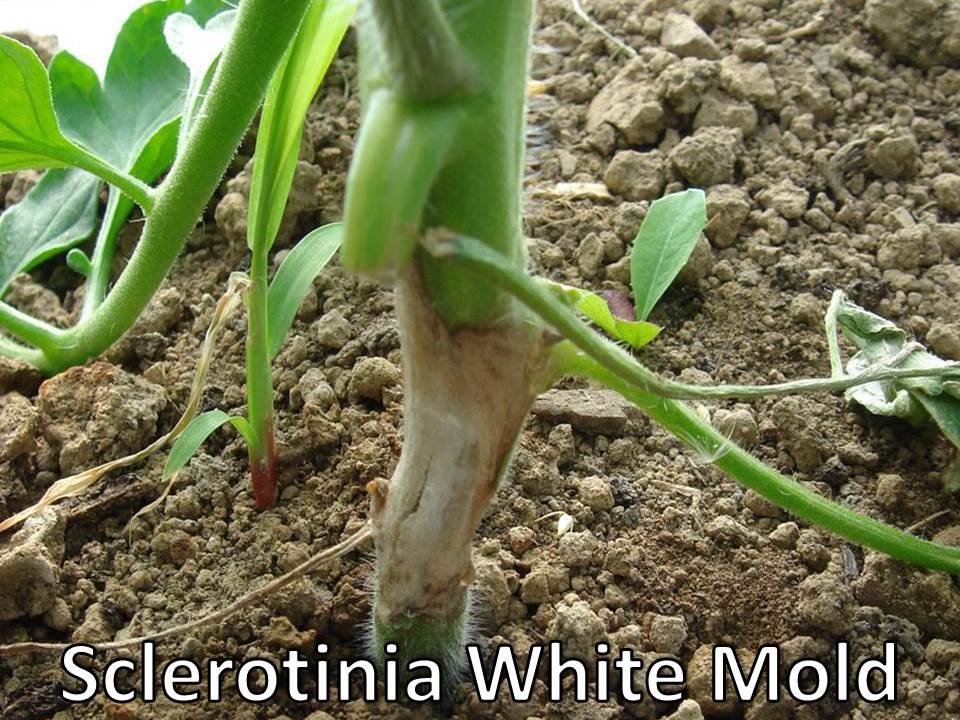
Initial infection of the pathogen is on tissues within the plant canopy, often near the base of the stem at the soil line. Bleached areas and watery soft rots form on the stems and leaf axils and then wet, fluffy white mold develops inside and outside the plant tissue. The white cottony mycelium spreads to the stems, leaves, petioles, and flowers. As the disease symptoms become more severe, the plant wilts and infected tissues become further bleached and brittle. In 7 to 10 days, clumps of mycelium form black sclerotia that are white-pinkish inside. The sclerotia are easily spread by equipment, plant debris, workers, and water.
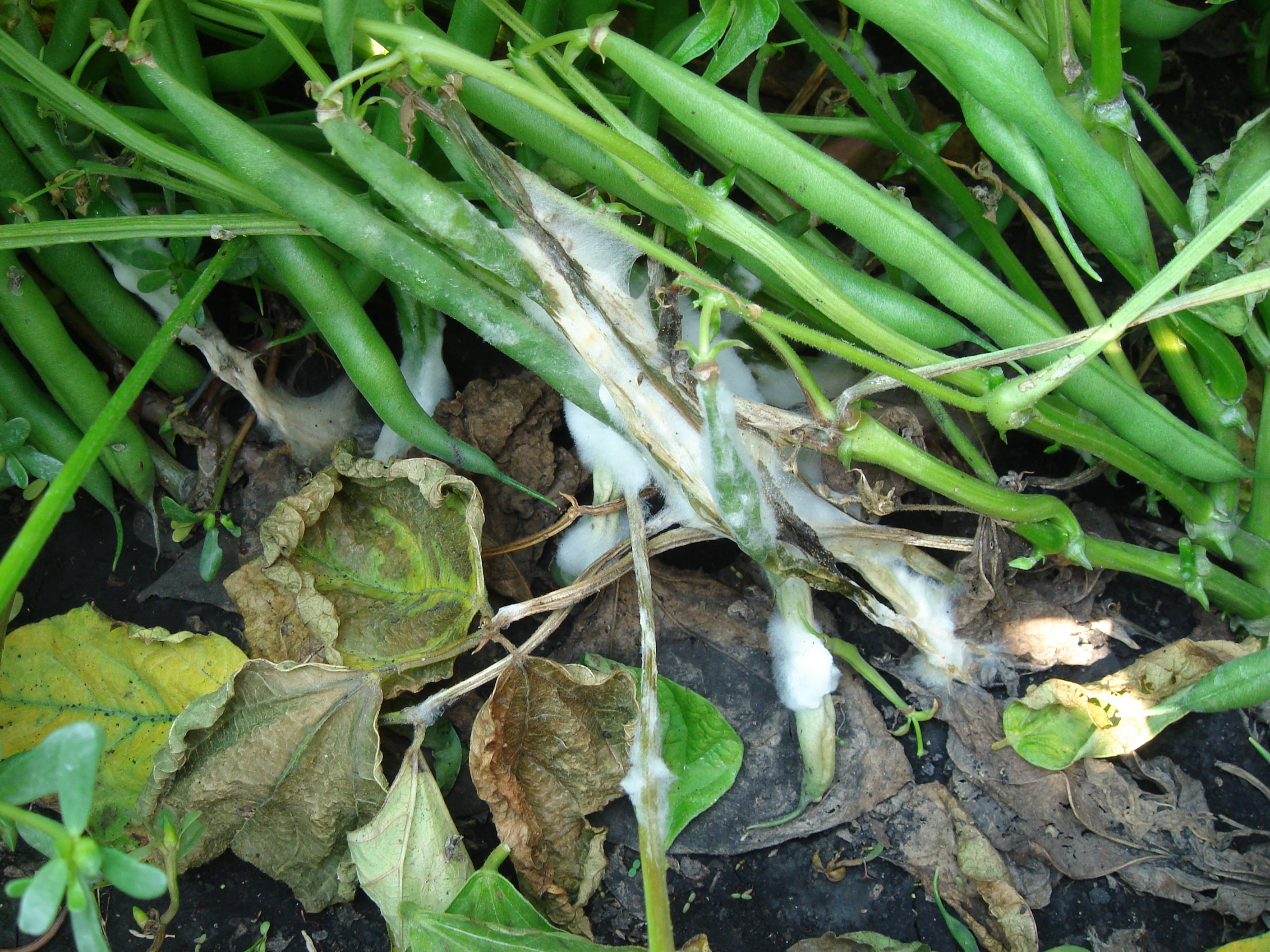

Sclerotinia white mold on bean plants (left), tomato plants (center), and pepper plants (right).
Signs
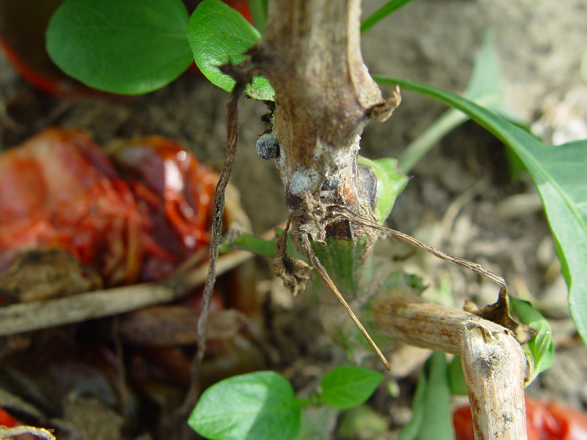
Signs of S. sclerotiorum are visible white mycelial masses and black sclerotia with white-pink centers. Apothecia formed from sclerotia near the soil line may be visible.


An image of black sclerotia formed in the stem of a tomato plant (left) and an image of apothecia germinating in soil (right).
Often Confused With
- Botrytis grey mold: A grey fuzzy spore-producing mycelium grows on all infected plant parts including buds, leaves, flowers, and fruit when conditions are humid and warm.

Isolation Media
To isolate Sclerotinia sclerotiorum, collect the sclerotia from host plant tissue or from the soil. To isolate from soil, a sieve with 0.43 – 2.00mm openings is required. All sclerotia should be surface-sterilized with NaOCl or CaOCl to prevent contamination by secondary pathogens. Allow the sclerotia to dry before re-germinating them directly on moist filter paper or incubating them in plastic bags with moist filter paper. The hyphae can be plated onto PDA media. Refer to the NC State University’s webpage on Sclerotinia sclerotiorum for more information on isolating the pathogen from sclerotia.
- A semi-selective blue medium is used to trap Sclerotinia sclerotiorum ascospores, where the medium will change from blue to yellow when a high number of ascospores are trapped.
- A semi-selective medium is used to isolate Sclerotinia sclerotiorum from seed.
Rapid Diagnostic Tests
- End-point Polymerase Chain Reaction (PCR) Assay
- Discriminatory simplex and multiplex PCR for four species of the genus Sclerotinia, Abd-Elmagid et. al. 2013
- Sclerotinia sclerotiorum Primers
- SSaspr F/ SSaspr R
- Sclerotinia minor
- SMLcc2 F/ SMLcc2 R
- Sclerotinia trifoliorum
- STCad F/ STCad R
- Sclerotinia sclerotiorum Primers